Pneumonia. Pneumonia is an inflammation of the lungs of an infectious nature involving all structural elements of the lung tissue and obligatory damage to the lung tissue
ACUTE PNEUMONIA Acute pneumonia is a group of acute infectious inflammatory diseases of the lungs, different in etiology, pathogenesis and morphological characteristics, with predominant damage to the respiratory sections and the presence of intra-alveolar exudate. Most often caused by bacteria, mycoplasmas and viruses. According to clinical and morphological features, lobar (lobar) pneumonia, bronchopneumonia (focal) and interstitial pneumonia are distinguished.
LOUPIC PNEUMONIA There are the following synonyms that reflect the morphological features of lung damage: lobar, fibrinous, pleuropneumonia. Lobar pneumonia is an infectious-allergic disease. It is an independent nosological form. The causative agent is pneumococcus types 1, 2 and 3, rarely - Klebsiella (Friedlander's diplobacillus). In the pathogenesis, an immediate hypersensitivity reaction is of great importance. Characteristically, the alveoli of the entire lobe are affected simultaneously while the bronchi remain intact. Always accompanied by fibrinous pleurisy (pleuropneumonia).
Stages of lobar pneumonia. 2. Red liver stage. 2nd day. Microscopic picture: the alveoli are filled with exudate, consisting of fibrin and erythrocytes. Macroscopic picture: the affected lobe is enlarged, dense (hepatic), red, fibrinous deposits on the pleura (fibrinous pleurisy).
Stages of lobar pneumonia. 3. Stage of gray hepatization. 4 -6 days. Microscopic picture: the capillaries are empty, in the alveolar exudate there is fibrin, leukocytes, macrophages, fibrinous deposits on the pleura. Macroscopic picture: the affected lobe is enlarged, dense, granular in section, uniform in appearance, gray in color.
COMPLICATIONS OF PNEUMONIA 1. Pulmonary. A. Carnification (organization of exudate in the lumen of the alveoli). b. Lung abscess. V. Gangrene (wet). 2. Extrapulmonary. Occur when the infection spreads lymphogenously or hematogenously. Includes purulent mediastinitis, pericarditis, peritonitis, purulent arthritis, acute ulcerative endocarditis (usually tricuspid valve), purulent meningitis, brain abscess.
COMPLICATIONS OF PNEUMONIA Pathomorphosis. It manifests itself as loss of one or another stage and abortive forms, a decrease in the frequency of complications. Causes of death. The mortality rate is about 3%. Death occurs from acute cardiopulmonary failure or purulent complications.
COMPLICATIONS OF PNEUMONIA Lobar Friedlander pneumonia. More often it occurs as a nosocomial (nosocomial) infection. Old people, newborns and alcoholics are sick. Characterized by necrosis of the alveolar septa with frequent formation of abscesses, foci of carnification and severe interstitial fibrosis.
BRONCHOPNEUMONIA (FOCAL PNEUMONIA) Makes up the bulk of acute pneumonia. Polyetiological. The most common pathogens are bacteria: pneumococci, staphylococci, streptococci, Pseudomonas aeruginosa, etc. It can occur as a nosocomial infection in weakened patients, and is usually caused by gram-negative microorganisms (Klebsiella, Pseudomonas aeruginosa and Escherichia coli) and Staphylococcus aureus.
BRONCHOPNEUMONIA More often occurs as an autoinfection. Depending on the characteristics of the pathogenesis, autoinfectious bronchopneumonia can be aspiration, hypostatic, postoperative, and also developing against the background of immunodeficiency. More often it is a complication of other diseases. Bronchopneumonia of newborns and the elderly, as well as some etiological variants of bronchopneumonia (for example, legionella) can be considered as independent nosological forms.
BRONCHOPNEUMONIA Morphological manifestations. The bronchi are initially affected. Inflammation of the alveoli spreads from the bronchial wall in a descending manner in case of endobronchitis or peribronchially in case of panbronchitis or destructive bronchiolitis. Exudate can be serous, purulent, hemorrhagic, mixed. According to the prevalence of the process, acinar, lobular, confluent lobular, segmental, and miliary pneumonia are distinguished.
Features of some common bacterial bronchopneumonia a. Pneumococcal pneumonia. It is more common in elderly and debilitated patients, especially with cardiopulmonary pathology (hypostatic pneumonia). Often complicated by pleural empyema.
STAPHYLOCOCCAL PNEUMONIA Staphylococcal pneumonia (Staphylococcus aureus). Usually occurs as a complication of respiratory viral infections (flu, etc.). It often develops in drug addicts with intravenous infection, as well as in weakened elderly patients with chronic pulmonary diseases. Typically abscessed, the development of pleural empyema often serves as a source of septicopyemia.
Pneumonia caused by Pseudomonas aeruginosa. Pneumonia caused by Pseudomonas aeruginosa. One of the most common nosocomial infections. Abscess formation and pleurisy are characteristic. When the infection is hematogenously introduced into the lungs (usually from extensive festering wounds), coagulation necrosis and a hemorrhagic component are characteristic. The prognosis is bad.
INTERSTITIAL PNEUMONIA Inflammation develops mainly in the alveolar septa with secondary accumulation of exudate in the lumens of the alveoli. Synonyms: alveolitis, pneumonitis. The process may be diffuse or limited. Caused by certain pathogens: viruses, fungi, mycoplasmas, chlamydia (ornithosis), rickettsia (Q fever-pneumorickettsiosis), pneumocysts.
VIRAL PNEUMONIA a. Viral pneumonia. Most common in childhood. More often caused by influenza viruses, parainfluenza, respiratory syncytial virus, adenovirus (see “Airborne infections”). Hyperplasia of the alveolar epithelium is characteristic with the formation of giant cells that differ in appearance in different diseases; squamous metaplasia of the bronchiolar epithelium is possible. Often complicated by secondary bacterial infection.
VIRAL PNEUMONIA The most common viral pneumonia in immunodeficiency states is cytomegalovirus pneumonia (opportunistic infection). It is characterized by predominantly mononuclear infiltration of the alveolar septa, hyperplasia of the alveolar epithelium, the appearance of large cells with characteristic intranuclear inclusions, and serous fluid in the lumens of the alveoli
Mycoplasma pneumonia. Also known as “atypical pneumonia”. One of the most common forms of non-bacterial pneumonia. Usually occurs in children and adolescents. The onset is more inconspicuous and erased than with bacterial pneumonia. It is characterized by an inflammatory lymphoplasmacytic infiltrate of the alveolar septa, hyperplasia of the alveolar epithelium, the presence of intra-alveolar hyaline membranes, exudate in the lumen of the alveoli may be absent, but is often combined with changes characteristic of bronchopneumonia: the appearance of leukocytes in the lumen of bronchioles and alveoli.
Pneumocystis pneumonia. An opportunistic infection most common in patients with HIV infection. It also occurs in other forms of immunodeficiency. Caused by P. carinii, an opportunistic microorganism classified as a protozoan (some classify it as a fungus). In persons with impaired cellular immunity, it can develop due to the previous presence of pneumocystis in the pulmonary foci of latent infection or as a result of fresh infection
Pneumocystis pneumonia. Characterized by desquamation of alveolar epithelial cells and filling of the alveoli with foamy fluid containing pneumocystis, as well as plethora and lymphohistiocytic infiltration of the alveolar septa with possible destruction of them. Characterized by increasing shortness of breath against the background of mild physical and radiological signs. It can occur in the form of a mixed infection with the addition of other flora (fungi, cytomegalovirus, cocci, mycobacteria, etc.).
The work can be used for lessons and reports on the subject "Philosophy"
In this section of the site you can download ready-made presentations on philosophy and philosophical sciences. The finished presentation on philosophy contains illustrations, photographs, diagrams, tables and the main theses of the topic being studied. A philosophy presentation is a good method of presenting complex material in a visual way. Our collection of ready-made presentations on philosophy covers all philosophical topics of the educational process both at school and at university.
Lecturer:Professor, Doctor of Medical Sciences Nedelskaya S.N.
Lecture outline
1.2.
3.
4.
5.
6.
Relevance
Etiology
Pathogenesis
Diagnostic principles
Principles of treatment and prevention
Conclusion
Pneumonia -
Pneumonia, acute infectious inflammatorylung disease
with availability
intraalveolar
exudation
ICD code J12-J18
Classification of pneumonia
According to the conditions of occurrence1. Out-of-hospital (home).
2. Intrahospital (hospital, nosocomial) – develops
after 48 hours of hospital stay or 48 hours after
discharge from the hospital.
3. Pneumonia of newborns (neonatal):
a) intrauterine (congenital) – developed in the first 72 hours
child's life
b) acquired (postnatal)
Community-acquired
In-hospital
4. Ventilation (in children on mechanical ventilation)
a) early – up to 72 hours on mechanical ventilation
b) late – after 4 or more days of mechanical ventilation
5. Aspiration
6. Pneumonia in persons with immunodeficiency conditions.
Classification of pneumonia
According to clinical and radiological data:1. Focal
3. Croupous (lobar)
2. Segmental
4. Interstitial
Localization:
1. One-sided (right, left)
2. Double-sided
Flow:
1. Acute (up to 6 weeks)
2. Prolonged (more than 6 weeks – up to 8 months)
3. Recurrent
Severity
l ll
lll
lV
V
Respiratory failure
I Art.
II Art.
III Art.
Classification of pneumonia
Complications1. Uncomplicated
2. Complicated
GENERAL VIOLATIONS
Toxic-septic condition
ITS
Cardiovascular syndrome
DIC syndrome
Changes in the central nervous system
PULMONARY
PROCESS
Destruction
Abscess
Pleurisy
Pneumothorax
INFLAMMATION
DIFFERENT
ORGANS
Sinusitis
Otitis
Pyelonephritis
Meningitis
osteomyelitis
Order of the Ministry of Health of Ukraine dated 01/04/12-8-1178
from 12/14/2009
Pneumonia severity scale in children
Degreegravity
Quantity
points
Risk of mortality
Rendering
help
l
<50
0,1
Outpatient
ll
51-70
0,6
Outpatient
lll
71- 90
2,8
Hospitalization
lV
91-130
8,2
Hospitalization
V
>130
29,2
Hospitalization
Pneumonia severity index in children
SignsPoints
Age
Tachycardia
+10
Up to 6 months
+25
Leukocytosis
+10
6 months-3 years
+15
Leukopenia
+20
3-15 years
+10
Anemia
+10
Cyanosis
+15
Concomitant pathology
Innate. heart defects
+30
pH<7,35
+30
Hypotrophy
+10
BUN (residual nitrogen) >11 mmol/l
+20
Kidney pathology
+10
Hct<30%
+10
Unodeficient state
+10
Sa O2< 90%
+20
CVC(cardiovascular disorders)
+20
Impaired consciousness
Dyspnea
+20
Multilobar infiltration on
X-ray
+15
Toxic encephalopathy
+30
ITS
+40
Chest pain
+10
Destruction
+50
Body temperature >39°C or<36°С
+15
Pleural exudate
+30
Anatomical and physiological characteristics of the child’s body that predispose to the development of pneumonia
The trachea and large bronchi are short and wide - penetrates easilyinfection
Small bronchi and bronchioles are narrow, easily collapsed and
become obstructed
Features of bronchial branching - frequent involvement in
pathological process of I, II, IX, X, VI segments of both lungs and IV, V
left lung segments
Lack of elastic elements and surfactant, tendency to
development of atelectasis and emphysema
Insufficient mucociliary clearance - difficulty in removal
foreign particles
Insufficient synthesis of interferons and immunoglobulins (Ig A) –
inferior immune response
The pulmonary parenchyma is plethoric, well vascularized,
rich in interstitium, at birth it is in a compressed
condition
Scheme of the segmental structure of the lungs
Risk factors
PrematuritySevere perinatal pathology: intrauterine
hypoxia, asphyxia, birth trauma
Syndrome of cyclic vomiting, regurgitation
Artificial feeding
Rickets
Anemia
Hypotrophy
Congenital heart defects
Cystic fibrosis
Lung malformations
Surgical interventions
Hereditary immunodeficiencies
Hypovitaminosis
Chronic foci of infection of the ENT organs
Smoking
Etiology of pneumonia in children
AgePathogens
newborns
Streptococci group B, Enterobacteriaceae (E.coli
and etc.)
1-3 months
Viruses, Enterobacteriaceae (E.coli, etc.),
H.influenzae, C.trachomatis, S.aureus
3 months – 5 years
Viruses, S. pneumoniae, H. influenzae.
More than 5 years
S. pneumoniae, M. pneumoniae,
Ch.Pneumoniae Mycoplasma, Chlamydia
pneumoniae
Pathogens of pneumonia in children depending on the conditions of infection
Out-of-hospitalNaya
Pneumococcus
Wand
influenza
In-hospital
Staphylococcus
Intestinal
wand
Wand
influenza
Staphylococcus
Bronhamella
Streptococcus Proteus
Pseudomonas
Mycoplasma
Serrations
Chlamydia
Mycoplasma
pneumonia
Enterobacter
Legionella
Klebsiella
Anaerobes
Perinatal For immunodeficiencies
Chlamydia
trichomonas
Pneumocysts
Ureaplasma
Cytomegalovirus
Bronhamella
Streptococcus
Bacteroides
Listeria
All bacteria and
cocci
Pneumocysts
Cytomegalovirus
Mycobacteria
Mushrooms
Main phases of pathogenesis
Phase Basic pathogenetic processes1
Penetration of microorganisms and edema-inflammatory obstruction of the upper respiratory tract
2
Processes of nonspecific inflammation
3
Activation of free radical processes
oxidation
4
Violation of pathophysiological mechanisms
breathing regulation
5
Respiratory failure and impairment
non-respiratory lung functions
6
Metabolic and functional disorders
organs and systems
Diagnostic criteria: clinic
Fever (above 38 oC)Toxicosis
Dyspnea
Respiratory failure
Cough
Cyanosis
Physical symptoms – shortening
percussion sound, weakened
breath, different-sized wet
wheezing, crepitus
Respiratory failure
A condition in which the lungs are unable toprovide normal gas
blood composition, which leads to a decrease
functional capabilities of the body,
As a result, hypoxemia develops and
hypercapnia
Characteristics of respiratory failure in pneumonia
DEGREEClinical characteristics
External
breath
Gas composition
blood
1
Shortness of breath during physical activity, cyanosis MOD increased,
perioral for anxiety, vital capacity and blood volume
P:D=2.5:1, tachycardia, blood pressure is normally reduced
Little changed
2
Shortness of breath at rest, constant,
perioral cyanosis, face and hands
- constant. Blood pressure is increased.
Tachycardia.
P:D=2-1.5:1
MOD increased,
Saturation
Vital capacity decreased
blood O2 70 more than 25-85%, acidosis
30%
3
Severe shortness of breath (heart rate more than
150% of the norm),
generalized cyanosis.
Blood pressure is reduced.
P:D varies
MOD, VC and DO
reduced more
than 50%
Saturation
blood O2 is lower
70%,
decompensated
acidosis
Diagnostic criteria
Laboratory research:- Leukocytosis, increased ESR, neutrophilia
- Increase in C - RB, seromucoids
The absence of hematological changes does not allow us to exclude
pneumonia!!!
X-ray examination – “Golden”
diagnostic standard
- homogeneous shadows of various densities and sizes
- cloud-like shadows, disseminated changes (with
interstitial pneumonia)
- focal-confluent, dense with a bulging border (with
destruction)
- basal changes (with viral pneumonia)
- atelectasis (with prolonged course)
Clinical diagnostic algorithm
STARTINSPECTION:
T more than 38 oС
more than 3 days
and/or retractions
chest
cells (without
phenomena
bronchial obstruction)
Local
systems:
shortening
percussion
sound and/or
weakened
or
bronchial
breath
and/or
local
wheezing
X-ray or start of treatment
Asymmetry
wet
wheezing
Toxicosis,
Leukocytosis
higher
15*109/l Purchased
community-acquired (typical and
atypical)
Pneumonia with
immunodeficient
states Hospital (nosocomial) Elements of destructive
pneumonia
Subtotal blackout
destruction
Bullous pneumonia Destruction (tomogram) Mycoplasma
pneumonia
Legionella
pneumonia
Chlamydia
pneumoniae
Differential diagnosis
ARVIAcute bronchitis
Bronchiolitis
Bronchial asthma
Foreign body aspiration
Chronic lung diseases
Tuberculosis
Features of the course in young children
Depends on gender (more often boys), age (thanThe smaller the child, the more severe the disease),
premorbid state, pathogen
Develops against the background of ARVI, measles, whooping cough
Percussion changes are minimal,
auscultation – variety of respiratory
noise
In newborns due to apnea or bradypnea
NPV may be increased slightly,
there may not be hyperthermia
Pneumococcal pneumonia
At the age of 3-7 years it proceeds asfocal
Over the age of 7 years it causes
lobar pneumonia
High fever up to 39-40 oC,
“rusty” sputum, DN.
Features of pneumonia caused by staphylococcus
70% young childrenpreceded by staphylococcal infections
(pyoderma, otitis, conjunctivitis, etc.)
One-sided process - often on the right,
can be bilateral
Intoxication is pronounced
Bright percussion and auscultation
painting
Progresses quickly
Pulmonary and extrapulmonary complications,
abscess formation, presence of bullae
Bacteremia 20 – 50% of cases
The prognosis is serious
Features of pneumonia caused by streptococcus
Streptococcal infection (tracheitis,regional lymphadenitis, lymphangitis
Focal, focal-confluent pneumonia
Protracted current
Purulent complications (damage to the pleura,
abscesses, osteomyelitis, etc.)
Bacteremia up to 10%
Onset gradual or acute
Severe intoxication, fever
Physical data from minimal to
fine wheezing
Recovery is slow – 1-2 months
Features of pneumonia caused by Haemophilus influenzae
Caused in association with pneumococcus orvirus
In children with impaired immunity,
age-related immune crisis conditions
systems.
Accompanying epiglotitis, laryngotracheitis
Gradual start
The process is often bilateral
Possible destruction of interalveolar
septum, fibroplastic proliferation
Cough without phlegm
High fever
Leukocytosis, moderately increased ESR
Features of pneumonia caused by chlamydia
Chlamydia pneumoniaeOften pharyngitis, fever, enlarged cervical
lymph nodes
A week later, wheezing in the lungs and shortening
sound
Interstitial foci of infiltration on
X-ray
The condition is not serious
Blood is not inflammatory
Chlamydia trachomatis
Source – mother’s birth canal
Preceded by conjunctivitis
Most often between 3 – 19 weeks of life
Persistent cough, no fever
intoxication, eosinophilia
Significant infiltrative changes on
X-ray
Indications for hospitalization
Children under 3 years oldComplicated course of the disease
Respiratory failure and unstable
hemodynamics
Children with malnutrition
Children with congenital heart defects,
tracheobronchial tree and lungs
Concomitant chronic diseases
Unfavorable social and living conditions
General principles of therapy
Bed restVentilation of the room (access
fresh air), oxygen therapy
(humidified with oxygen)
Detoxification – drinking plenty of fluids,
according to indications - infusion therapy
Antibiotic therapy
Pathogenetic therapy
Symptomatic therapy 2 PRINCIPLES FOR CHOOSING ANTIBIOTIC
FOR TREATING PNEUMONIA:
EMPIRICAL (APPROXIMATE) -
ANTIBIOTIC PRESCRIPTION BEFORE IDENTIFICATION
MICROORGANISMS THAT CAUSE PNEUMONIA.
THERAPY IS BASED ON ASSUMPTION
POSSIBLE PATHOGEN USED AS A STARTER
FOR OUT-OF-HOSPITAL (HOME) PNEUMONIA.
ETIOLOGICAL (EXACT) –
PRESCRIPTION OF AN ANTIBIOTIC AFTER ETIOLOGICAL
PATIENT IDENTIFICATION WITH DEFINITION
SENSITIVITY TO ANTIBIOTIC.
A step-by-step approach to antibiotic therapy for pneumonia!!!
Drugs of choice:Penicillins and semisynthetic penicillins
Cephalosporins 1st and 2nd generations
Macrolides
Lincosamines
Reserve drugs:
Antistaphylococcal and antipseudomonas penicillins
Parenteral CF 2 and 3 generations
Aminoglycosides 2 and 3 generations
Rifampicin
Fluoroquinolones
Deep reserve drugs:
Carbapenems
Monobactams
Vancomycin
Aminopenicillins and cephalosparins are used as initial therapy. In severe cases of pneumonia, a combination of these with ammo is prescribed.
As initial therapyuse aminopenicillins and
cephalosparins.
In severe cases of pneumonia -
prescribe their combination with
aminoglycosides
If you have a history of allergies to
penicillin - used
macrolides Antibiotic dosing
β-lactam aminopenicillins:
10-20 mg/kg per dose, with an interval of 8 hours
daily dose - 30-60 mg/kg/day
Cephalosparins: 50-100 mg/kg/day – daily dose for 2
introduction
Macrolides:
Clarithromycin 15 mg/kg/day – daily dose for 2 doses
Aminoglycosides:
Amikacin 15 mg/kg/day – daily dose for 2 administrations
Netilmicin 6-7.5 mg/kg/day daily dose for 2-3 administrations
Carbopenems 30-60 mg/kg/day – daily dose for 3 administrations
In severe cases, use parenteral
intravenous administration of antibiotics with transition to
Criteria for the effectiveness of antibiotic therapy for pneumonia
Efficiency assessment is carried out whenuncomplicated form of pneumonia after 24-48
hours, in case of complications after 48-72 hours
Criteria:
Dynamics of the child’s general condition
Dynamics of feverish reaction
Dynamics of respiratory rate and pulse and their
ratios
Dynamics of laboratory and radiological
data
Options for the effectiveness of antibiotic therapy
The full effect is a decrease in temperature of less than38 oC after 24-48 hours uncomplicated and after 72 hours with
complicated against the background of improved condition and appetite,
reducing shortness of breath
Partial – maintaining temperature above 38 oC
while reducing toxicosis, improving appetite and
absence of negative x-ray
paintings
No effect - maintaining temperature
above 38 oC with deterioration and/or increase
changes in the lungs or pleural cavity
Forecast
In the absence of complications and concomitant diseases– favorable
With a favorable course of the disease, pneumonic
the lesion resolves before the end of 3-4 weeks (with prolonged
course of pneumonia with incomplete resorption is possible
formation of a focus of pneumosclerosis and chronicity of the process)
Discharge of the patient to a children's institution - no earlier than 2
weeks from the onset of the disease with persistent clinical
picture of recovery, data normalization
laboratory and x-ray examination
Children who have had lobar pneumonia are observed in
on an outpatient basis in the recovery room
treatment for 3 (children under 3 years of age) – 2
months (children over 3 years old)
Dispensary observation is carried out for 8 months
– 1 year (includes examination by a local doctor,
dynamic blood tests)
Slide 2
Pneumonia (ancient Greek πνευμονία from πνεύμων) (pneumonia) - inflammation of the lung tissue, usually of infectious origin with predominant damage to the alveoli
Slide 3
Slide 4
The term “pneumonia” unites a large group of diseases, each of which has its own etiology, pathogenesis, clinical picture, radiological signs, characteristic laboratory findings and treatment features. It can occur as an independent disease or as a complication of other diseases.
Slide 5
The main diagnostic method is x-ray examination of the lungs
Slide 6
The main method of treatment is antibacterial therapy.
- Antibiotics are the cornerstone of treatment for pneumonia. The choice of antibiotic depends on the microorganism that caused the pneumonia.
- Drugs that dilate the bronchi and dilute sputum are also used - orally or in the form of inhalations, corticosteroids, intravenous saline solutions, oxygen.
- Sometimes pleural puncture and bronchoscopy are performed.
- Physiotherapy is often used: ultraviolet therapy, vibration massage, exercise therapy, paraffin, ozokerite
Slide 7
- Late diagnosis and delay in starting antibacterial therapy (more than 8 hours) worsen the prognosis of the disease.
- Possible death.
- Serious complications of pneumonia may include: abscess and gangrene of the lung, pleurisy, pleural empyema, obstruction, acute respiratory failure, endocarditis, pericarditis, meningitis, pulmonary edema, sepsis
Slide 8
picture of a healthy person's lungs
Slide 9
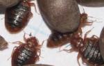
photo of the lungs of a person suffering from pneumonia
Slide 10
Pneumonia may be
- focal - i.e. occupy a small focus of the lung (bronchopneumonia - respiratory sections + bronchi);
- segmental - spread to one or more segments of the lung;
- lobar - to capture a lobe of the lung. A classic example of lobar pneumonia is lobar pneumonia - mainly the alveoli and the adjacent area of the pleura;
- confluent - merging of small foci into larger ones;
- total - pneumonia is called if it spreads to the entire lung.
In addition, pneumonia may be
Unilateral, if only one lung is affected,
Bilateral, if both lungs are sick.
Slide 11
- The incidence of pneumonia depends on many factors: standard of living, social and family status, working conditions, contact with animals, travel, bad habits, contact with sick people, individual characteristics of a person, and the geographic distribution of a particular pathogen.
- Pneumonia remains one of the most common causes of death in children and elderly people in our time, especially in social institutions (orphanages, boarding schools, prisons).
- The incidence of pneumonia in elderly patients increases sharply while they are being treated in hospitals for another disease.
View all slides
Pneumonia is an acute infectious (mainly bacterial) disease, an acute infectious (mainly bacterial) disease, characterized by focal lesions of the respiratory parts of the lungs with intra-alveolar exudation, detected during physical and radiological examinations, characterized by focal lesions of the respiratory parts of the lungs with intra-alveolar exudation, detected during physical and radiological examinations examinations, accompanied by feverish reactions and intoxication expressed to varying degrees, accompanied by feverish reactions and intoxication expressed to varying degrees

Classification of pneumonia Community-acquired Typical (without pronounced immune disorders) In patients with severe immunodeficiency conditions of various origins Aspiration (lung abscess) Nosocomial Actually nosocomial Ventilator-associated Early VAP Late VAP Nosocomial pneumonia in patients with severe IDS Associated with the provision of medical care Pneumonia in residents of nursing homes Other categories of patients (ABT in the previous 3 months, hospitalization for more than 2 days in the previous 90 days, stay in long-term care facilities, etc.)

Community-acquired pneumonia is an acute disease, an acute disease that arose in a community setting, or diagnosed in the first 48 hours from the moment of hospitalization, arose in a community setting, or diagnosed in the first 48 hours from the moment of hospitalization, accompanied by symptoms of lower respiratory tract infection (fever, cough, sputum production, chest pain, shortness of breath, etc.) and accompanied by symptoms of lower respiratory tract infection (fever, cough, sputum production, chest pain, shortness of breath, etc.) and radiological signs of “fresh” focal infiltrative changes in the lungs by radiological signs of “fresh” focal infiltrative changes in the lungs in the absence of an obvious diagnostic alternative in the absence of an obvious diagnostic alternative

Nosocomial pneumonia - pneumonia that develops in a patient no earlier than 48 hours from the moment of hospitalization, not caused by an infection that was in the incubation period at the time of admission; pneumonia that develops in a patient no earlier than 48 hours from the moment of hospitalization, not caused by an infection, was in the incubation period at the time of admission

Pneumonia associated with the provision of medical care has been separately identified in recent years in foreign literature; according to the conditions of occurrence, they are community-acquired, however, the SPECTRUM OF PATIENTS AND THEIR ANTIBIOTIC RESISTANCE PROFILE is similar to the pathogens of nosocomial pneumonia; according to the conditions of occurrence, they are community-acquired, however, THE SPECTRUM OF PATIENTS AND THEIR ANTIBIOTIC RESISTANCE PROFILE is similar to pathogens of nosocomial pneumonia

Epidemiology of pneumonia The incidence of pneumonia in the Republic of Belarus averages 10.0-13.8 per 1000 population, increasing among people over 50 years of age to 17.0 per 1000 population The incidence of pneumonia in the Republic of Belarus averages 10.0-13.8 per 1000 population , increasing among people over 50 years of age to 17.0 per 1000 population in the United States, about 4 million cases of community-acquired pneumonia are registered annually in the United States, about 4 million cases of community-acquired pneumonia are registered annually in the United States, the cost of treating CAP in the USA is about 10 billion dollars per year the cost of treating VAP in the USA is about 10 billion dollars per year, pneumonia ranks first in the structure of mortality from infectious diseases in the USA, pneumonia ranks first in the structure of mortality from infectious diseases in the USA


The main pathogenetic mechanism for the development of pneumonia: aspiration of oropharyngeal secretion with colonizing microorganisms contained in it (relevant for S.pneumoniae, H.influenzae, Gr-bacteria, anaerobes) aspiration of oropharyngeal secretion with colonizing microorganisms contained in it (relevant for S.pneumoniae, H. influenzae, Gr-bacteria, anaerobes) microaspiration (often) microaspiration (often) macroaspiration (rarely in the presence of predisposing factors - stroke, chronic alcoholism, repeated vomiting) macroaspiration (rarely in the presence of predisposing factors - stroke, chronic alcoholism, repeated vomiting)

Other more rare pathogenetic mechanisms for the development of pneumonia: Inhalation of microbial aerosol (Mycoplasma pneumoniae, Chlamydophila pneumoniae, Legionella pneumoniae, etc.) Inhalation of microbial aerosol (Mycoplasma pneumoniae, Chlamydophila pneumoniae, Legionella pneumoniae, etc.) Hematogenous dissemination from the extrapulmonary focus of infection Hematogenous dissemination from the extrapulmonary focus of infection Direct spread of infection from adjacent foci of pathology (intrahepatic or subphrenic abscess, etc.) Direct spread of infection from adjacent foci of pathology (intrahepatic or subphrenic abscess, etc.) Reactivation of latent infection (Pneumocystis jiroveci in the case of severe IDS) Reactivation latent infection (Pneumocystis jiroveci in case of severe IDS)

Etiology of community-acquired pneumonia (CAP): 1. Streptococcus pneumoniae – “king of CAP”, 30-50% of all cases 2. Atypical microorganisms (Chlamydophila pneumoniae, Mycoplasma pneumoniae, Legionella pneumophila) – up to 30% of all cases 3. Other rare pathogens of CAP – 3-5% of all cases: 1. Haemophilus influenzae 2. Staphylococcus aureus 3. Klebsiella pneumoniae, even less often - other Gr-bacteria of the Enterobacteriaceae family

The etiology of CAP is determined by a number of factors: the age of the patients, the severity of the disease, the presence of concomitant pathology (risk factors), etc. The etiology of CAP is determined by a number of factors: the age of the patients, the severity of the disease, the presence of concomitant pathology (risk factors), etc. In adults undergoing CAP, a mixed infection is often detected (in one clinical trial, almost every second of 346 examined patients with pneumococcal etiology of the disease showed serological signs of active infection caused by mycoplasmas or chlamydia). In adults undergoing CAP, a mixed infection is often detected (in one In clinical trials, almost every second of the 346 examined patients with pneumococcal etiology of the disease showed serological signs of active infection caused by mycoplasmas or chlamydia)

The main causative agents of CAP in outpatients: mild CAP in persons under 60 years of age without concomitant pathology; non-severe CAP in persons over 60 years of age and/or with concomitant pathology S.pneumoniae M. pneumoniae C.pneumoniae S.pneumoniae H.influenzae C.pneumoniae S. aureus Enterobacteriaceae

The main causative agents of CAP in hospitalized patients: non-severe CAP – hospitalization in the general hospital; severe CAP – hospitalization in the intensive care unit S.pneumoniae H.influenzae C.pneumoniae S. aureus Enterobacteriaceae S.pneumoniae Legionella spp. S.aureus Enterobacteriaceae

Risk factors and possible causative agents of CAP alcoholism: S.pneumoniae, anaerobes, Gr-bacteria (usually K.pneumoniae) alcoholism: S.pneumoniae, anaerobes, Gr-bacteria (usually K.pneumoniae) COPD/smoking: S.pneumoniae, H. influenzae, M. catarrhalis, Legionella spp. COPD/smoking: S.pneumoniae, H.influenzae, M.catarrhalis, Legionella spp. decompensated diabetes: S.pneumoniae, S.aureus decompensated diabetes: S.pneumoniae, S.aureus stay in nursing homes: S.pneumoniae, representatives of the family Enterobacteriaceae, H.influenzae, S.aureus, C. pneumoniae, anaerobes stay in nursing homes: S.pneumoniae, representatives of the family Enterobacteriaceae, H.influenzae, S.aureus, C. pneumoniae, anaerobes

Risk factors and possible causative agents of CAP influenza epidemic: S.pneumoniae, S.aureus, S.pyogenes, H.influenzae influenza epidemic: S.pneumoniae, S.aureus, S.pyogenes, H.influenzae CAP development against the background of bronchiectasis, cystic fibrosis: P.aeruginosa, B.cepacia, S.aureus development of PAP against the background of bronchiectasis, cystic fibrosis: P.aeruginosa, B.cepacia, S.aureus contact with air conditioners, air humidifiers, water cooling systems: L.pneumophila contact with air conditioners, air humidifiers , water cooling systems: L.pneumophila unsanitized oral cavity, expected massive aspiration: anaerobes unsanitized oral cavity, expected massive aspiration: anaerobes

“Pyramid” of lower respiratory tract infections (Macfarlane J.T. Lower respiratory tract infection and pneumoniae in the community) Died (1-2) Hospitalized in the ICU (1-2) Hospitalized for pneumonia (20) Diagnosed with community-acquired pneumonia (100) Persons those who received antibiotics (2,000) Those who sought medical help (8,000 patients) Persons with symptoms of community-acquired LRTIs (patients)

Diagnosis of pneumonia - subjective complaints: suspicion of pneumonia should arise with fever in combination with complaints of cough, shortness of breath, sputum production, chest pain; suspicion of pneumonia should arise with fever in combination with complaints of cough, shortness of breath, sputum production, chest pain cage in elderly patients, respiratory complaints may be absent, and in the clinic symptoms of a general nature will prevail: drowsiness during the day and insomnia at night, confusion, fatigue, heavy sweating at night, nausea, vomiting, signs of exacerbation or decompensation of concomitant diseases of internal organs in elderly patients, respiratory complaints may be absent, and in the clinic symptoms of a general nature will prevail: drowsiness during the day and insomnia at night, confusion, fatigue, heavy sweating at night, nausea, vomiting, signs of exacerbation or decompensation of concomitant diseases of internal organs

Diagnosis of pneumonia - objective data classic objective signs of pneumonia: classic objective signs of pneumonia: shortening of the percussion tone over the affected area of the lung shortening of the percussion tone over the affected area of the lung locally auscultated bronchial breathing locally auscultated bronchial breathing focus of sonorous fine-bubble rales or crepitus focus of sonorous fine-bubble rales or crepitus increased bronchophony and vocal tremor increased bronchophony and vocal tremor in interstitial pneumonia, characterized by the presence of dry and moist rales without signs of compaction of the lung tissue; interstitial pneumonia is characterized by the presence of dry and moist rales without signs of compaction of the lung tissue; in 20% of patients, objective signs of PAP may differ from typical ones or be absent in general, in 20% of patients, objective signs of PFS may differ from typical ones or be absent altogether

Diagnosis of pneumonia - instrumental examination almost always requires a chest x-ray to confirm the diagnosis, because Numerous studies have shown the low sensitivity and specificity of an objective clinical examination in the diagnosis of CAP; almost always, a chest x-ray is required to confirm the diagnosis, because Numerous studies have shown the low sensitivity and specificity of an objective clinical examination in the diagnosis of CAP in typical cases of CAP; the diagnosis criterion is the detection of focally infiltrative or interstitial changes in the lungs in typical cases of CAP; the diagnosis criterion is the detection of focally infiltrative or interstitial changes in the lungs; in some cases, changes in X-ray may be absent despite the presence of clinical and physical signs of pneumonia; in some cases, changes on the X-ray may be absent despite the presence of clinical and physical signs of pneumonia

Possible causes of clinical and radiological dissociation: deep neutropenia with the impossibility of developing a localized acute inflammatory reaction in the lung tissue; deep neutropenia with the impossibility of developing a localized acute inflammatory reaction in the lung tissue; early stages of the disease (according to stetoacoustic data, pneumonia can be recognized an hour before the appearance of pulmonary infiltrate on an x-ray) early stages of the disease (according to stetoacoustic data, pneumonia can be recognized an hour before the appearance of pulmonary infiltrate on a radiograph) in the case of Pneumocystis pneumonia in HIV-infected patients, pathological changes on the radiograph are absent in 10-20% of patients in the case of Pneumocystis pneumonia in HIV-infected patients, pathological changes on the radiograph absent in 10-20% of patients. In case of doubt, in the presence of obvious clinical symptoms of pneumonia and the absence of changes on the radiograph, computed tomography is indicated (the most sensitive for detecting interstitial changes in the lungs)

Diagnosis of pneumonia - microbiological examination The material most often is freely coughed up sputum The material most often is freely coughed up sputum The effectiveness of a microbiological examination depends on the rules for collecting the material (optimally before the start of antibacterial therapy) and the conditions of its transportation The effectiveness of a microbiological examination depends on the rules for collecting the material (optimally before the start of antibacterial therapy) and conditions of its transportation at the first stage of the study, sputum is Gram stained; if there are less than 25 polymorphonuclear leukocytes and more than 10 epithelial cells (when viewing at least 10 fields of view with a magnification of X 100), cultural examination is not advisable, because the sample is contaminated with the contents of the oral cavity; at the first stage of the study, the sputum is Gram stained; if there are less than 25 polymorphonuclear leukocytes and more than 10 epithelial cells (when viewing at least 10 fields of view with a magnification of X 100), cultural examination is not advisable, because the sample is contaminated with oral contents

Diagnosis of pneumonia - microbiological examination sputum microscopy can provide guidelines when choosing antibacterial therapy (lanceolate Gr+ diplococci - S.pneumoniae, weakly stained Gr-coccobacilli - H.influenzae, etc.) sputum microscopy can provide guidelines when choosing antibacterial therapy (lanceolate Gr+ diplococci – S.pneumoniae, weakly stained Gr- coccobacilli – H.influenzae, etc.) at the second stage of the study, sputum culture is carried out to isolate specific pathogens and determine the antibiotic resistance profile; at the second stage of the study, sputum culture is carried out to isolate specific pathogens and to determine the antibiotic resistance profile in seriously ill patients, before starting antibacterial therapy, it is also necessary to perform venous blood cultures (2 samples from 2 different veins, at least 10 ml of blood for each sample); in seriously ill patients, before starting antibacterial therapy, it is also necessary to perform venous blood cultures (2 samples from 2 different veins, at least 10 ml of blood for each sample)!!! Despite the importance of obtaining laboratory material before prescribing an antibiotic, microbiological testing should not be a reason for delaying antibacterial therapy (especially in severe patients)

Participation in the development of bronchopulmonary inflammation is not typical for a number of microorganisms: Streptococcus viridans Streptococcus viridans Staphylococcus epidermidis and other coagulase-negative staphylococci Staphylococcus epidermidis and other coagulase-negative staphylococci Enterococcus spp. Enterococcus spp. Neisseria spp. Neisseria spp. Candida spp. etc. Candida spp. etc. Isolation of this group of microorganisms from sputum indicates CONTAMINATION OF THE MATERIAL by the flora of the upper respiratory tract, and not the etiological significance of these pathogens in the development of pneumonia!!!

Diagnosis of pneumonia - laboratory data peripheral blood leukocytosis more than X 10 9 / L indicates a high probability of bacterial infection, and leukopenia below 3 X 10 9 / L or leukocytosis above 25 X 10 9 / L is an unfavorable prognostic sign peripheral blood leukocytosis more than X 10 9 /l indicates a high probability of bacterial infection, and leukopenia below 3 X 10 9 /l or leukocytosis above 25 X 10 9 /l is an unfavorable prognostic sign; detectable abnormalities in a biochemical blood test, indicating damage to certain organs/systems, have a prognostic value; detectable deviations in biochemical blood tests indicating damage to certain organs/systems have prognostic significance; a number of studies have shown that the highest concentration of C-reactive protein is observed in patients with severe pneumococcal or legionella pneumonia; a number of studies have shown that the highest concentration of C- reactive protein is observed in patients with severe pneumococcal or Legionella pneumonia

Diagnosis criteria The diagnosis of PAP is determined if the patient has radiologically confirmed focal infiltration of the lung tissue and at least two clinical signs from the following: acute fever at the onset of the disease above 38 *C acute fever at the onset of the disease above 38 *C cough with sputum cough with sputum physical signs (focus of crepitus and/or fine-bubble rales, harsh bronchial breathing, shortening of percussion sound) physical signs (focus of crepitation and/or fine-bubble rales, harsh bronchial breathing, shortening of percussion sound) leukocytosis above 10 X 10 9 /l and/or band shift (above 10%) leukocytosis above 10 X 10 9 /l and/or band shift (above 10%)

Choosing a place of treatment for patients with VBP, dividing patients with VBP into inpatient and outpatient is fundamentally important due to different approaches to diagnostic examination and tactics of antimicrobial chemotherapy; dividing patients with VBP into inpatient and outpatient is fundamentally important due to different approaches to diagnostic examination and tactics of antimicrobial chemotherapy 30-50 % of patients hospitalized for CAP are characterized as prognostically “favorable” and can be successfully treated at home 30-50% of patients hospitalized for CAP are characterized as prognostically “favorable” and can be successfully treated at home at present there are a number of clinical and laboratory scales, which, based on the assessment of the severity of VBP and prognosis, give recommendations on the choice of place of treatment; currently, there are a number of clinical and laboratory scales, which, based on the assessment of the severity of VBP and prognosis, give recommendations on the choice of place of treatment

Prognostic scale CRB-65 1C (Confusion) Impaired consciousness 2R (Respiratory rate) Respiratory rate (RR) equal to or higher than 30/min 3B (Blood pressure) Low diastolic or systolic blood pressure: 

Management of patients with CAP in an outpatient setting Diagnostic minimum: medical history medical history physical examination of the patient physical examination of the patient chest radiography optimally in two projections chest radiography optimally in two projections general blood count general blood count


Based on clinical, hematological, radiological data and using generally accepted microbiological methods, as a rule, it is impossible to reliably establish the etiology of PAP. The empirical choice of antibiotics, based on the most likely sensitivity of the most likely pathogens, forms the basis of therapy

Antibiotic resistance of the main causative agents of CAP S.pneumoniae – the main problem is resistance to beta-lactams and macrolides S.pneumoniae – the main problem is resistance to beta-lactams and macrolides multidrug-resistant pneumococcus – S.pneumoniae, resistant to three or more classes of antibiotics multidrug-resistant pneumococcus – S .pneumoniae, resistant to three or more classes of antibiotics pneumococcal resistance to penicillin is usually accompanied by co-resistance to I-II generation cephalosporins, tetracyclines and co-trimoxazole pneumococcal resistance to penicillin is usually accompanied by co-resistance to I-II generation cephalosporins, tetracyclines and co-trimoxazole

Antibiotic resistance of the main causative agents of PEGAS (year) - a multicenter study of antibiotic resistance of pneumococcus in Russia: PEGAS (year) - a multicenter study of antibiotic resistance of pneumococcus in Russia: the level of resistance to penicillin does not exceed 10%, while most strains are moderately resistant the level of resistance to penicillin does not exceed 10%, while most strains are moderately resistant; the level of resistance to CA III (ceftriaxone, cefotaxime) is not higher than 2%; the level of resistance to CA III (ceftriaxone, cefotaxime) is not higher than 2%; resistance to 14- and 15 -membered macrolides (erythromycin, clarithromycin, azithromycin) 6-9%, to 16-membered macrolides (josamycin, spiramycin, midecamycin) and lincosamides does not exceed 4.5% resistance to 14- and 15-membered macrolides (erythromycin, clarithromycin, azithromycin) 6-9%, to 16-membered (josamycin, spiramycin, midecamycin) and lincosamides does not exceed 4.5%

Antibiotic resistance of the main pathogens of PEGAS (year) - a multicenter study of antibiotic resistance of pneumococcus in Russia: PEGAS (year) - a multicenter study of antibiotic resistance of pneumococcus in Russia: high resistance of pneumococcus is characteristic of co-trimoxazole (40.7% of strains are insensitive) and tetracyclines (29.6% of strains are insensitive) high resistance of pneumococcus is characteristic of co-trimoxazole (40.7% of strains are insensitive) and tetracyclines (29.6% of strains are insensitive) co-trimoxazole and tetracyclines should not be used as drugs of choice for empirical therapy of CAP due to the high resistance of the main causative agent to them, co-trimoxazole and tetracyclines should not be used as drugs of choice for empirical treatment of CAP due to the high resistance of the main causative agent to them

Antibiotic resistance of the main causative agents of PEGAS (year) - a multicenter study of antibiotic resistance of pneumococcus in Russia: PEGAS (year) - a multicenter study of antibiotic resistance of pneumococcus in Russia: resistance of pneumococcus to: resistance of pneumococcus to: respiratory fluoroquinolones (levofloxacin) is not registered , moxifloxacin) respiratory fluoroquinolones (levofloxacin, moxifloxacin) vancomycin vancomycin linezolid linezolid resistance to chloramphenicol (levomycetin) does not exceed 8.6% resistance to chloramphenicol (levomycetin) does not exceed 8.6% resistance to amoxicillin does not exceed 0.5%, to amoxi cillin clavulanate - 0.3% resistance to amoxicillin does not exceed 0.5%, to amoxicillin clavulanate - 0.3% all penicillin-resistant pneumococci retained 100% sensitivity to amoxicillin clavulanate all penicillin-resistant pneumococci retained 100% sensitivity to amoxicillin clavulanate

Antibiotic resistance of the main causative agents of VBP PEGAS II (year) - a multicenter study of antibiotic resistance of H. influenzae in Russia: PEGAS II (year) - a multicenter study of antibiotic resistance of H. influenzae in Russia: the main mechanism of resistance is the production of beta-lactamases that hydrolyze aminopenicillins the main mechanism of resistance is the production of beta-lactamases that hydrolyze aminopenicillins; resistance to aminopenicillins did not exceed 4.7%; resistance to aminopenicillins did not exceed 4.7%; no strains resistant to amoxicillin clavulanate, cephalosporins II-IV, carbapenems, fluoroquinolones were identified; resistant to amoxicillin clavulanate, cephalosporins II-IV, carbapenems, fluoroquinolones


Group I - patients with non-severe CAP under the age of 60 years without concomitant pathology, the most common pathogens: S.pneumoniae, M.pneumoniae, C.pneumoniae drugs of choice: amoxicillin or macrolide antibiotics (clarithromycin, azithromycin) by mouth alternative drugs: respiratory fluoroquinolones (levofloxacin , gemifloxacin, moxifloxacin) orally!!! Despite the fact that in vitro aminopenicillins are not active against “atypical” pathogens of CAP, clinical studies have not revealed differences in the effectiveness of these antibiotics, as well as individual representatives of the class of macrolides or respiratory fluoroquinolones, the spectrum of action of which includes both typical and “ atypical" pathogens

Group II – patients with non-severe CAP 60 years of age and older and/or with concomitant diseases and risk factors Chronic diseases and risk factors influencing the etiology and prognosis of CAP: chronic obstructive pulmonary disease chronic obstructive pulmonary disease diabetes mellitus diabetes mellitus congestive heart failure congestive heart disease failure chronic renal failure chronic renal failure cirrhosis of the liver cirrhosis of the liver alcoholism, drug addiction alcoholism, drug addiction body weight deficiency body weight deficiency

Group II – patients with non-severe CAP 60 years of age and older and/or with concomitant diseases and risk factors, the most common pathogens: S.pneumoniae, H.influenzae, C.pneumoniae, S.aureus, family Enterobacteriaceae drugs of choice: combination therapy amoxicillin /clavulanate or amoxicillin/sulbactam orally + macrolide (azithromycin, clarithromycin) orally alternative drugs: monotherapy with respiratory fluoroquinolones (levofloxacin, moxifloxacin, gemifloxacin) orally

Drug Average doses (for adults) Ampicillin 1.0-2.0 g IV or IM every 6 hours Amoxicillin 0.5-1.0 g orally every 8 hours Amoxicillin/clavulanate 0.625 g orally every 6-8 hours 1, 2 g IV every 6-8 hours Cefuroxime 0.75-1.5 g IV, IM every 8 hours Cefuroxime axetil 0.5 g orally every 12 hours Cefotaxime 1.0-2.0 g IV, IV /m every 8 hours Ceftriaxone 1.0-2.0 g IV, IM every 24 hours Clarithromycin 0.5 g orally every 12 hours 0.5 g IV every 12 hours Azithromycin 3-day course: 0.5 g orally every 24 hours 5-day course: 0.5 g orally on the first day, then 0.25 g every 24 hours Midecamycin 0.4 g orally every 8 hours Levofloxacin 0.5 g orally every 24 hours 0.5 g IV every 24 hours Moxifloxacin 0.4 g orally and IV every 24 hours AB, often used in outpatient practice

Management of patients with CAP in an outpatient setting parenteral blockers in the treatment of CAP on an outpatient basis do not have proven advantages over oral parenteral blockers in the treatment of CAP on an outpatient basis do not have proven advantages over oral parenteral blockers should be used only in cases of expected low compliance when taking oral medications or when refusing hospitalization or if it is impossible to carry it out in a timely manner, parenteral antibiotics should be used only in cases of expected low compliance when taking oral medications or in case of refusal of hospitalization or the impossibility of carrying it out in a timely manner; the initial assessment of the effectiveness of therapy should be carried out within an hour from the start of therapy (efficacy criteria: reduction in temperature, reduction in symptoms of intoxication, etc. . clinical manifestations of the disease), the initial assessment of the effectiveness of therapy should be carried out within an hour from the start of therapy (efficacy criteria: decrease in temperature, reduction of symptoms of intoxication and other clinical manifestations of the disease) if treatment is ineffective, the tactics of antibacterial therapy should be reconsidered and the advisability of hospitalization of the patient should be re-evaluated if treatment is ineffective it is necessary to reconsider the tactics of antibacterial therapy and re-evaluate the advisability of hospitalization of the patient in case of non-severe CAP, the average duration of ABT is 7-10 days (ABT is completed with stable normalization of body temperature for 3-4 days) in case of non-severe CAP, the average duration of ABT is 7-10 days (ABT is completed when stable normalization of body temperature within 3-4 days)

In hospitalized patients, a more severe course of CAP is implied, so it is advisable to start therapy with parenteral antibiotics. In hospitalized patients, a more severe course of CAP is implied, so it is advisable to start therapy with parenteral antibiotics after 3-4 days of treatment when body temperature normalizes, intoxication and other symptoms of the disease decrease. switching from parenteral to oral use of AB before completing the full course of therapy (stepped therapy); after 3-4 days of treatment, with normalization of body temperature, reduction of intoxication and other symptoms of the disease, it is possible to switch from parenteral to oral use of AB before completing the full course of therapy (stepped therapy) !!! In case of severe CAP, the appointment of antibiotics should be urgent - a delay in their appointment of 4 hours or more significantly worsens the prognosis of the disease, increases mortality and length of hospital stay (Houck P.M et al. Timing of antibiotic administration and outcomes for Medicare patients hospitalized with community-acquired pneumoniae Clin Infect Dis 2003;36:)

Group I – non-severe CAP in hospitalized patients most common pathogens: S.pneumoniae, H.influenzae, C.pneumoniae, S.aureus, family Enterobacteriaceae drugs of choice: combination therapy benzylpenicillin IV, IM ± macrolide orally benzylpenicillin IV /in, intramuscular ± macrolide inside ampicillin intravenous, intramuscular ± macrolide inside ampicillin intravenous, intramuscular ± macrolide inside amoxicillin/clavulanate intravenously ± macrolide inside amoxicillin/clavulanate intravenous ± macrolide inside cefuroxime IV, IM ± macrolide orally Cefuroxime IV, IM ± macrolide orally Cefotaxime or ceftriaxone IV, IM ± macrolide orally Cefotaxime or ceftriaxone IV, IM ± macrolide orally According to series studies, the presence in the initial treatment regimen of a drug active against “atypical” microorganisms improves the prognosis and reduces the length of stay of patients in the hospital, which makes the use of combination therapy beta-lactam + macrolide justified alternative drugs: monotherapy respiratory fluoroquinolones (levofloxacin, moxifloxacin) i.v. respiratory fluoroquinolones (levofloxacin, moxifloxacin) IV azithromycin IV (can be used as monotherapy only in the absence of risk factors for antibiotic-resistant pneumococci, Gr-enterobacteria and infection caused by Ps.aeruginosa) azithromycin IV (can be used as monotherapy only in the absence of risk factors for antibiotic-resistant pneumococci, Gr-enterobacteria and infection caused by Ps.aeruginosa)

Group II – severe PAP in hospitalized patients, the most common pathogens: S.pneumoniae, Legionella spp., S.aureus, family Enterobacteriaceae drugs of choice: combination therapy amoxicillin/clavulanate IV + macrolide IV amoxicillin/clavulanate IV + macrolide IV cefotaxime IV + macrolide IV cefotaxime IV + macrolide IV ceftriaxone IV + macrolide IV ceftriaxone IV + macrolide IV alternative drugs: combination therapy respiratory fluoroquinolones (levofloxacin , moxifloxacin) IV + III generation cephalosporins IV respiratory fluoroquinolones (levofloxacin, moxifloxacin) IV + III generation cephalosporins IV In the presence of risk factors for infection caused by Pseudomonas aeruginosa (bronchiectasis, taking systemic corticosteroids, therapy with broad-spectrum antibiotics effects for more than 7 days during the last month, exhaustion) drugs of choice are ceftazidime, cefepime, cefoperazone/sulbactam, ticarcillin/clavulanate, piperacillin/tazobactam, carbapenems (meropenem, imipenem)

Management of patients with CAP in inpatient conditions, the initial assessment of the effectiveness of therapy should be carried out 48 hours after the start of treatment, and in the case of severe disease - 24 hours later (efficacy criteria: reduction in temperature, reduction of symptoms of intoxication and respiratory failure) the initial assessment of the effectiveness of therapy should be carried out 48 hours after the start of treatment, and in case of severe disease - 24 hours later (efficacy criteria: reduction in temperature, reduction in symptoms of intoxication and respiratory failure) if treatment is ineffective (maintenance of high fever and intoxication or progression of disease symptoms), the tactics of antibacterial therapy should be reconsidered if treatment is ineffective (persistence of high fever and intoxication or progression of disease symptoms), the tactics of antibacterial therapy should be reconsidered for non-severe CAP; the average duration of ABT is 7-10 days (ABT is completed with persistent normalization of body temperature within 3-4 days); for non-severe CAP, the average duration of ABT 7-10 days (ABT ends with persistent normalization of body temperature within 3-4 days) for severe PAP, a 10-day course of ABT is recommended for severe PAP, a 10-day course of ABT is recommended if there is evidence of mycoplasma or chlamydial etiology for PAP, antibacterial therapy is extended until 14 days if there is evidence of a mycoplasma or chlamydial etiology for CAP, antibacterial therapy is extended to 14 days for CAP of staphylococcal etiology or CAP caused by gram-negative enterobacteria, as well as for legionella CAP, the duration of ABT should be from 14 to 21 days for CAP of staphylococcal etiology or CAP, caused by gram-negative enterobacteria, as well as with legionella CAP, the duration of ABT should be from 14 to 21 days

Typical errors in antibiotic therapy for CAP: errors in choosing a drug aminoglycosides - gentamicin and other aminoglycosides are inactive against pneumococcus and atypical microorganisms aminoglycosides - gentamicin and other aminoglycosides are inactive against pneumococcus and atypical microorganisms ampicillin orally - has low bioavailability when taken orally, amoxicillin is used orally ampicillin orally – has low bioavailability when taken orally, amoxicillin is used orally; first generation cephalosporins (cefazolin, etc.) are inactive against most pathogens of respiratory infections, and are inferior in antipneumococcal activity to aminopenicillins and most cephalosporins of later generations; penicillin-resistant pneumococci are cross-resistant to first generation cephalosporins; activity against H. influenzae is not clinically significant; sensitive to beta-lactamases, which produce almost 100% of M. catarrhalis strains. First generation cephalosporins (cefazolin, etc.) are inactive against most pathogens of respiratory infections, and are inferior in antipneumococcal activity to aminopenicillins and most cephalosporins of later generations; penicillin-resistant pneumococci are cross-resistant to first generation cephalosporins; activity against H. influenzae is not clinically significant; sensitive to beta-lactamases, which are produced by almost 100% of M. catarrhalis strains co-trimoxazole - high resistance to this drug S. pneumoniae and H. influenzae, frequent allergic skin reactions, the presence of safer drugs co-trimoxazole - high resistance to this drug S.pneumoniae and H.influenzae, frequent skin allergic reactions, the presence of safer drugs ciprofloxacin and other fluoroquinolones of the second generation - has low activity against S.pneumoniae and atypical pathogens; if used rashly, it forms resistance to fluoroquinolones of all generations, including respiratory ciprofloxacin and other fluoroquinolones of the second generation - has low activity against S.pneumoniae and atypical pathogens; if used rashly, it forms resistance to fluoroquinolones of all generations, including respiratory ones

Typical mistakes in antibiotic therapy for CAP: late start of antibacterial therapy: prescribing antibiotics later than 4 hours after the diagnosis of community-acquired pneumonia leads to an increase in mortality; late start of antibacterial therapy: prescribing antibiotics later than 4 hours after the diagnosis of community-acquired pneumonia leads to an increase in mortality; frequent changes of antimicrobial medications during treatment; “explained” by the danger of developing resistance, frequent changes in AMPs during treatment, “explained” by the danger of developing resistance. There are clear indications for replacing AMPs: clinical ineffectiveness, which can be judged after an hour of therapy; clinical ineffectiveness, which can be judged after an hour of therapy; development of serious adverse reactions; requiring discontinuation of AMPs development of serious adverse reactions requiring discontinuation of AMPs high potential toxicity of AMPs (for example, aminoglycosides), limiting the duration of their use high potential toxicity of AMPs (for example, aminoglycosides), limiting the duration of their use continuation of antibiotic therapy while certain radiological and/or laboratory changes persist until until their complete disappearance, continuation of antibiotic therapy while maintaining individual radiological and/or laboratory changes until their complete disappearance. The main criterion for discontinuing antibiotics is regression of clinical symptoms. The persistence of individual laboratory and/or radiological changes is not an absolute indication for continued antibiotic therapy

Prolonged PFS in the majority of patients with PFS, by the end of 3-5 days after the start of effective ABT, body temperature normalizes and other clinical manifestations of the disease regress, while radiological recovery, as a rule, lags behind the clinical one in the majority of patients with PFS by the end of 3-5 days after the start of effective ABT normalizes body temperature and regresses other clinical manifestations of the disease, while radiological recovery, as a rule, lags behind the clinical one if, against the background of an improvement in the clinical picture, by the end of the 4th week from the onset of the disease it is not possible to achieve complete radiological resolution of focal infiltrative changes in the lungs, we should talk about protracted or non-resolving (slowly resolving) PFS if, against the background of an improvement in the clinical picture, by the end of the 4th week from the onset of the disease it is not possible to achieve complete radiological resolution of focal infiltrative changes in the lungs, we should talk about protracted or non-resolving (slowly resolving) PFS

Risk factors for the development of prolonged CAP: age over 65 years age over 65 years alcoholism alcoholism presence of concomitant disabling diseases of internal organs (COPD, congestive heart failure, renal failure, malignant neoplasms, diabetes mellitus, etc.) presence of concomitant disabling diseases of internal organs (COPD, congestive heart failure, renal failure, malignant neoplasms, diabetes mellitus, etc.) severe CAP severe CAP multilobar infiltration multilobar infiltration highly virulent pathogens (L.pneumophila, S.aureus, gram-negative enterobacteria) highly virulent pathogens (L.pneumophila, S .aureus, gram-negative enterobacteria) smoking smoking clinical ineffectiveness of initial therapy (persisting leukocytosis and fever) clinical ineffectiveness of initial therapy (persisting leukocytosis and fever) secondary bacteremia secondary bacteremia secondary resistance of pathogens to antibiotics secondary resistance of pathogens to antibiotics

Algorithm for examining a patient with protracted community-acquired pneumonia syndrome: in the presence of risk factors for a protracted course of the disease, a control radiographic examination after 4 weeks. In the case of persistent pneumonic infiltration, an additional examination of the patient (CT, fibrobronchoscopy, etc.) is indicated in the presence of risk factors for a protracted course of the disease - a control radiographic examination after 4 weeks. In the case of persistent pneumonic infiltration, additional examination of the patient is indicated (CT, fibrobronchoscopy, etc.) in the absence of risk factors for a protracted course of the disease, the patient needs additional examination in the absence of risk factors for a protracted course of the disease, the patient needs additional examination

Compliance (adherence) - the patient’s agreement to follow the doctor’s recommendations Compliance (adherence) - the patient’s agreement to follow the doctor’s recommendations Incompliance - any deviation of the patient from medical instructions Incompliance - any deviation of the patient from medical instructions
Lecture plan Definition, relevance of pneumonia Definition, relevance of pneumonia Pathogenesis of pneumonia Pathogenesis of pneumonia Classification of pneumonia Classification of pneumonia Criteria for the diagnosis of pneumonia Criteria for the diagnosis of pneumonia Treatment principles: organization of the regime, aerotherapy, antibacterial therapy, immunotherapy and physiotherapeutic methods of treatment, prevention Principles of treatment: organization of the regime, aerotherapy, antibacterial therapy, immunotherapy and physiotherapeutic treatment methods, prevention

Pneumonia is a nonspecific inflammation of the lung tissue, which is based on infectious toxicosis, respiratory failure, water-electrolyte and other metabolic disorders with pathological changes in all organs and systems of the child’s body. Pneumonia is a nonspecific inflammation of the lung tissue, which is based on infectious toxicosis, respiratory failure, water-electrolyte and other metabolic disorders with pathological changes in all organs and systems of the child’s body.

Relevance: 1) The incidence of pneumonia ranges from 4 to 20 cases per 1000 children aged 1 month to 15 years. 2) In Ukraine, there has been an increase in the prevalence of pneumonia among children in the last three years (from 8.66 to 10.34). 3) Mortality from pneumonia among children in the first year of life is from 1.5 to 6 cases per child, which is 3-5% in the overall structure of mortality in children under 1 year of age. 4) Every year about 5 million children die from pneumonia in the world.

Etiology Hospital-acquired (nosocomial) pneumonia in most cases is caused by Ps. aeruginosa, also – Kl. pneumonia, St. aureus, Proteus spp. etc. These pathogens are resistant to antibiotics, which leads to severe disease and mortality. Hospital-acquired (nosocomial) pneumonia in most cases is caused by Ps. aeruginosa, also – Kl. pneumonia, St. aureus, Proteus spp. etc. These pathogens are resistant to antibiotics, which leads to severe disease and mortality. Community-acquired pneumonia (home, non-hospital). The spectrum of pathogens depends on the age of the patients. Community-acquired pneumonia (home, non-hospital). The spectrum of pathogens depends on the age of the patients.

Newborns: depends on the spectrum of urogenital infections in women. - Postnatal pneumonia is often caused by group B streptococci, less often by E. coli, Klebsiella pneumoniae, St. aureus, St. epidermalis. - Antenatal – streptococci of groups G, D, Ch. frachomatis, ureaplasma urealiticum, Listeria monocytogenes, Treponeta pallidum. - Children of the first half of the year: staphylococci, gram-negative intestinal flora, rarely - Moraxella catarrhalis, Str. рneumoniae, H. influenzae, Ch. trachomatis.

From 6 months to 5 years, Str. comes first. Pneumoniae (70-88% of all pneumonias) and H. influenzae type b (Hib infection) - up to 10%. These children also often isolate respiratory syncytial virus, influenza viruses, parainfluenza, rhino- and adenoviruses, but most authors consider them to be factors that contribute to infection of the lower respiratory tract with bacterial flora. From 6 months to 5 years, Str. comes first. Pneumoniae (70-88% of all pneumonias) and H. influenzae type b (Hib infection) - up to 10%. These children also often isolate respiratory syncytial virus, influenza viruses, parainfluenza, rhino- and adenoviruses, but most authors consider them to be factors that contribute to infection of the lower respiratory tract with bacterial flora.

In children 6-15 years old: bacterial pneumonia makes up % of all pneumonia and is caused by pneumococci Str. pyogenes; M. pneumoniae (23-44%), Ch. Pneumoniae (15-30%). The role of Hib infection decreases. In children 6-15 years old: bacterial pneumonia makes up % of all pneumonia and is caused by pneumococci Str. pyogenes; M. pneumoniae (23-44%), Ch. Pneumoniae (15-30%). The role of Hib infection decreases. In case of insufficiency of humoral immunity, pneumococcal, staphylococcal, and cytomegalovirus pneumonia are observed. In case of insufficiency of humoral immunity, pneumococcal, staphylococcal, and cytomegalovirus pneumonia are observed. In case of primary cellular immunodeficiencies, with long-term glucocorticoid therapy - P. carinii, M. avium, fungi of the genus Candida, Aspergilus. Often viral-bacterial and bacterial-fungal associations (65-80%). In case of primary cellular immunodeficiencies, with long-term glucocorticoid therapy - P. carinii, M. avium, fungi of the genus Candida, Aspergilus. Often viral-bacterial and bacterial-fungal associations (65-80%).

Pathogenesis In the pathogenesis of the development of acute pneumonia, V.G. Maidannik distinguishes six phases. In the pathogenesis of the development of acute pneumonia, V.G. Maidannik distinguishes six phases. The first is contamination by microorganisms and edematous-inflammatory destruction of the upper respiratory tract, dysfunction of the ciliated epithelium, and spread of the pathogen along the tracheobronchial tree. The first is contamination by microorganisms and edematous-inflammatory destruction of the upper respiratory tract, dysfunction of the ciliated epithelium, and spread of the pathogen along the tracheobronchial tree. The second is the primary alteration of lung tissue, activation of lipid peroxidation processes, and development of inflammation. The second is the primary alteration of lung tissue, activation of lipid peroxidation processes, and development of inflammation. Third: damage by prooxidants not only to the structures of the pathogen, but also to the macroorganism (surfactant); destabilization of cell membranes; phase of secondary toxic autoaggression. The area of damage to lung tissue increases. Third: damage by prooxidants not only to the structures of the pathogen, but also to the macroorganism (surfactant); destabilization of cell membranes; phase of secondary toxic autoaggression. The area of damage to lung tissue increases.

Fourth: disruption of tissue respiration, central regulation of respiration, ventilation, gas exchange and perfusion of the lungs. Fourth: disruption of tissue respiration, central regulation of respiration, ventilation, gas exchange and perfusion of the lungs. Fifth: the development of DN and disruption of the non-respiratory function of the lungs (cleansing, immune, excretory, metabolic, etc.). Fifth: the development of DN and disruption of the non-respiratory function of the lungs (cleansing, immune, excretory, metabolic, etc.). Sixth: metabolic and functional disorders of other organs and systems of the body. The most severe metabolic disorders are observed in newborns and young children. Sixth: metabolic and functional disorders of other organs and systems of the body. The most severe metabolic disorders are observed in newborns and young children.

There are 4 ways of contamination of the lungs with pathogenic flora: There are 4 ways of contamination of the lungs with pathogenic flora: 1) aspiration of the contents of the oropharynx (microaspiration during sleep) - the main way; 2) airborne; 3) hematogenous spread of the pathogen from an extrapulmonary source of infection; 4) Spread of infection from adjacent tissues of neighboring organs.


Clinical criteria for pneumonia in a newborn child: aggravated ante- and intrapartum anamnesis; burdened ante- and intrapartum anamnesis; pallor, perioral and acrocyanosis; pallor, perioral and acrocyanosis; groaning breath; groaning breath; tension and swelling of the wings of the nose; retraction of the pliable areas of the chest; tension and swelling of the wings of the nose; retraction of the pliable areas of the chest; respiratory arrhythmia; respiratory arrhythmia; rapid increase in pulmonary heart failure and toxicosis; rapid increase in pulmonary heart failure and toxicosis;

Muscle hypotonia, inhibition of newborn reflexes; muscle hypotonia, inhibition of newborn reflexes; hepatolienal syndrome; hepatolienal syndrome; weight loss; weight loss; coughing; less frequent cough; coughing; less frequent cough;

Increased body temperature; may be normal in immature newborns; increased body temperature; may be normal in immature newborns; hemogram: neutrophilic leukocytosis, shift of the formula to the left; hemogram: neutrophilic leukocytosis, shift of the formula to the left; X-ray: pulmonary tissue infiltrates, usually on both sides; increased pulmonary pattern in perifocal areas. X-ray: pulmonary tissue infiltrates, usually on both sides; increased pulmonary pattern in perifocal areas.

Clinical criteria for the diagnosis of pneumonia in young children: wet or unproductive cough; wet or unproductive cough; shortness of breath, breathing with the participation of auxiliary muscles; shortness of breath, breathing with the participation of auxiliary muscles; distant wheezing in broncho-obstructive syndrome; distant wheezing in broncho-obstructive syndrome; general weakness, refusal to eat, delayed weight gain; general weakness, refusal to eat, delayed weight gain; pale skin, perioral cyanosis, worsens with exercise; pale skin, perioral cyanosis, worsens with exercise;

Violation of thermoregulation (hyper- or hypothermia, toxicosis); violation of thermoregulation (hyper- or hypothermia, toxicosis); hard bronchial or weakened breathing, after 3-5 days moist rales appear; hard bronchial or weakened breathing, after 3-5 days moist rales appear; shortening of percussion sound in the projection of infiltrate; shortening of percussion sound in the projection of infiltrate; hemogram: neutrophilic leukocytosis, shift of the formula to the left; hemogram: neutrophilic leukocytosis, shift of the formula to the left; X-ray: infiltrates of lung tissue, increased pulmonary pattern in perifocal areas. X-ray: infiltrates of lung tissue, increased pulmonary pattern in perifocal areas.

70 in 1 min. respiratory arrhythmia Retraction of the intercostal spaces is moderate + pronounced Participation of auxiliary muscles in the act of breathing - + pronounced Cyanosis" title=" Criteria for the degree of DN Clinical manifestations of the degree of DN IIIIII Tachyptic with load at rest >70 in 1 min respiratory arrhythmia Retraction of intercostal spaces moderate + pronounced Participation of auxiliary muscles in the act of breathing - + pronounced Cyanosis" class="link_thumb"> 34 !} Criteria for the degree of DN Clinical manifestations Degree of DN IIIIII Tachypneous with load at rest >70 in 1 min. respiratory arrhythmia Retraction of intercostal spaces moderate + pronounced Participation of auxiliary muscles in the act of breathing - + pronounced Cyanosis mild perioral perioral, pronounced widespread Other manifestations - - nodding of the head in rhythm with breathing 70 in 1 min. respiratory arrhythmia Retraction of the intercostal spaces is moderate + pronounced Participation of auxiliary muscles in the act of breathing - + pronounced Cyanosis "> 70 in 1 min. respiratory arrhythmia Retraction of the intercostal spaces moderate + pronounced Participation of auxiliary muscles in the act breathing - + pronounced Cyanosis mild perioral perioral, pronounced widespread Other manifestations - - nodding the head in time with breathing "> 70 in 1 min. respiratory arrhythmia Retraction of the intercostal spaces is moderate + pronounced Participation of auxiliary muscles in the act of breathing - + pronounced Cyanosis" title=" Criteria for the degree of DN Clinical manifestations of the degree of DN IIIIII Tachyptic with load at rest >70 in 1 min respiratory arrhythmia Retraction of intercostal spaces moderate + pronounced Participation of auxiliary muscles in the act of breathing - + pronounced Cyanosis"> title="Criteria for the degree of DN Clinical manifestations Degree of DN IIIIII Tachypneous with load at rest >70 in 1 min. respiratory arrhythmia Retraction of intercostal spaces moderate + pronounced Participation of auxiliary muscles in the act of breathing - + pronounced Cyanosis"> !}
Treatment of pneumonia Children with acute pneumonia can be treated at home or in a hospital. Indications for hospitalization are as follows: Children with acute pneumonia can be treated at home and in hospital. Indications for hospitalization are as follows: 1) vital indications – intensive care and resuscitation measures are required; 2) decreased reactivity of the child’s body, the threat of complications; 3) unfavorable living conditions of the family, there is no possibility of organizing a “hospital at home”.

In a hospital, the child should be in a separate room (box) to prevent cross-infection. Until the age of 6, the mother must be with the child. The room should be wet cleaned, quartzed, and ventilated (4-6 times a day). The room should be wet cleaned, quartzed, and ventilated (4-6 times a day). The head of the bed should be raised. The head of the bed should be raised.

Nutrition Depends on the age of the child. In a serious condition of a patient in the 1st year of life, the number of feedings can be increased by 1-2, while complementary feeding can be excluded for several days. The main food is breast milk or adapted milk formula. If oral rehydration is necessary, rehydron, gastrolit, ORS 200, herbal tea, in fractions are prescribed.

Treatment of respiratory failure 1) Ensure clear airways. 2) Microclimate of the room: fresh, fairly humid air, the temperature in the room should be 18-19ºС. 3) In case of stage 2 respiratory failure, oxygen therapy is added: through a nasal tube - % oxygen utilization; through a mask – %, in an incubator – %, in an oxygen tent – %. For grade III DN, artificial ventilation is required.

Antibacterial therapy Basic principles of rational antibacterial therapy in children. 1. Start of treatment - after diagnosis. It is advisable to conduct flora cultures to determine sensitivity to antibiotics. Results will be available in 3-5 days. We select initial therapy empirically, taking into account the patient’s age, home or hospital pneumonia, and regional characteristics. 2. First course – broad-spectrum antibiotics (mainly β-lactams) are prescribed. 3. The main course - (replacement of the empirically selected antibiotic) depends on the culture result or the clinical picture. 4. The choice of dose depends on the severity, age, body weight.

5. Choice of route of administration: in severe cases, it is predominantly administered parenterally. 6. Choosing the frequency of administration: it is necessary to create a constant concentration of the antibiotic in the body. 7. Selection of a rational combination: synergism is required, only bactericidal or only bacteriostatic. Drugs should not enhance each other's toxic effects. 8. Conditions for stopping treatment: no earlier than 3 days of normal temperature and general condition of the child. The accuracy of empirical therapy may be %. The accuracy of empirical therapy may be %.

Antibacterial therapy for community-acquired pneumonia Age, form Etiology antibiotics starting regimen alternative regimen 1-6 months, typical mild E.coli Staph.aureus et epiderm. Streptoc. Pneumon. H.Influanzae Respiratory viruses (RS, parainfluenza) Oral: Protected aminopenicillins: Amoxcillin/clavlanate Ampicillin/sulbactam Oral or parenteral: I-II generation cephalosporins (cefuroxime, cefadroxil, cefazolin) 1-6 months, typical complicated 1-6 months, typical complicated Pseudomonas aeruginosa, Enterobaceriaceae, Proteus spp, E.coli, Klebsiella pneumoniae, Staph. aureus Parenteral: III-IV generation cephalosporins (cefotaxime, ceftazidime, cefepime) or amoxcilin/clavulanate + aminoglycosides (amikacin, netilmicin) Parenteral: amoxcilin/clavulanate + aminoglycosides Linkosamines Carbapenems + aminoglycosides

1-6 months, atypical, uncomplicated Chlamidia tr., pn., Mycoplasma pneumoniae, less often Pneumocyst Oral: modern macrolides (spiramycin, rox andromycin, azithromycin, josamycin) Oral: sulfamethoxazole trimethoprim 1-6 months, atypical, complicated Chlamydia tr ., pn., Mycoplasma pneumoniae, Pneumocyst Pneumocyst Intravenous: Spiramycin, erythromycin Intravenous: Fluoroquinolones (for health reasons) 6 months-6 years, typical, uncomplicated Streptoc.pneumon Hemophilus infl. Viruses (RS, parainfluenza, influenza, adenoviruses). Oral: Amoxcilin/clavulanate or amoxicillin Oral or parenteral: I-II generation cephalosporins (cefadroxil, cefuroxime) Modern macrolides (spiramycin, roxithromycin, azithromycin, dalosamycin)