What is seminoma and how long can they live with a tumor with proper therapy? Details about testicular seminoma: causes, symptoms, stages and prognosis for life Symptoms of seminoma
One of the malignant neoplasms in the male body is testicular seminoma. The source for tumor development is germ cells.
Mostly men from 25 to 40 years old, as well as after 65 years, are affected. This type of tumor metastasizes quite quickly, so it is very important to identify the disease at the initial stage. The prognosis with timely treatment is favorable.
Causes
It is not completely known what exactly causes this disease. According to the scientific works of some researchers, one can only make assumptions about possible reasons. According to one version, it follows that testicular seminoma can be caused by any precancerous conditions in which cell mutation occurs. As soon as the body is exposed to adverse factors, these cells degenerate into cancer cells.
According to another version, the cause of seminoma may be a hereditary factor. As practice shows, this diagnosis is most often made to persons whose immediate relatives have suffered from a testicular tumor. In addition, even if the relationship is distant, the risk still exists, since the exact type of inheritance is not defined.
One of the most common causes of seminoma is cryptorchidism. This is a pathology in which the testicle does not descend into the scrotum. But not all experts see a connection between these diseases. Research in this area is still being actively carried out today, and some scientists have already managed to establish a cause-and-effect relationship.
 Our regular reader got rid of PROSTATITIS using an effective method. He tested it on himself - the result was 100% - complete relief from prostatitis. This is a natural remedy based on honey. We tested the method and decided to recommend it to you. The result is fast. EFFECTIVE METHOD.
 There are several other versions that are expressed, but in fact are quite dubious. This:
When the lymph nodes surrounding the abdominal aorta are affected by metastases, the patient begins to experience severe abdominal pain. Swelling of the legs may occur due to compression of the inferior vena cava. Over time, the ureters are also subject to compression. This leads to problems with urination.
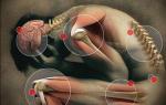
As the tumor grows, pain and heaviness appear in the testicle, as well as a feeling of discomfort in the lower abdomen
Subsequently, with the flow of lymph, metastases spread to the internal organs. As soon as the liver is affected, it enlarges and ascites rapidly develops. Penetration of metastases into the lungs causes a dry cough, accompanied by bronchospasm and shortness of breath, and the appearance of hemoptysis. Damage to the skeletal system causes severe pain and difficulty in movement.
The general condition of the patient is also affected. He becomes lethargic, loses interest in everything around him, becomes emotionally unstable, and feels general weakness. Loss of appetite and sudden weight loss occur. A depressive state with loss of ability to work often develops.
Diagnostics
The diagnosis is made based on external examination and additional examination methods. During visual inspection and palpation, asymmetry of the scrotum can be identified, in which a dense neoplasm can be clearly distinguished. If the patient suffers from cryptorchidism, the tumor is localized in the abdomen. Typically, the tumor does not cause discomfort upon palpation. Based on the accompanying symptoms, one can judge at what stage the disease is.
Instrumental diagnostics consists of ultrasound examination of the scrotum, chest x-ray, computed tomography of the abdominal organs, magnetic resonance imaging of the liver, and bone examination. In addition, a mandatory diagnostic step is a blood test for cancer markers.
An additional method of differential diagnosis can be aspiration biopsy, which allows identifying the tumor at the cellular level. If even after this type of diagnosis there are still doubts about the correct diagnosis, then an urgent operation is prescribed to remove the tumor, which is submitted for cytological examination. As soon as testicular seminoma is confirmed, additional research methods are prescribed to detect foci of metastasis.
Stages of seminoma
There are several stages of the disease, on which the further prognosis directly depends.
- At the first stage, only the testicle is involved in the pathological process. There are no metastases yet. This is the optimal time to fight the disease. If measures are not taken in time, metastases begin to spread.
- In the second stage, metastases begin to spread. Mostly, the retroperitoneal lymph nodes are the first to be affected. If treatment is started at this stage, in most cases it is possible to achieve complete recovery. But relapse occurs very often, so patients must undergo preventive examinations.
- At the third stage, metastases spread to organs that are located above the diaphragm. During this period, the patient is recommended to remain in a hospital setting. This will help track the dynamics of the disease and identify possible deteriorations.
- The fourth stage is the most severe, but it does not occur often, since already from the second stage the patient’s symptoms are quite pronounced, and he consults a doctor. The exception is a rapidly growing tumor, which for some reason is difficult to treat. At this stage, metastases have already spread to all lymph nodes and internal organs.
Testicular seminoma is one of these tumors, which makes itself felt very early. Already at the beginning of the second stage, the patient begins to experience pain, which becomes a reason to contact a specialist. And this is a big plus, as it helps to identify the disease when it has not yet been seriously advanced. In 90% of cases the prognosis is favorable.
But at the same time, this type of cancer is quite dangerous because it progresses very quickly, rapidly flowing from one stage to another. Therefore, therapy should be started immediately after diagnosis.
Chemotherapy
Treatment of seminoma
Treatment depends on the stage of the disease. This can be determined by conducting a complete examination. There are several common methods that are most often used to treat seminoma, namely:
- radiation therapy;
- chemotherapy;
- surgery.
It is important to remember that successful antitumor therapy can help completely get rid of the problem, but at the same time. Therefore, if the patient plans to have children in the future, then before starting the course of treatment he needs to save sperm samples in a specialized fertility center.
At an early stage of the disease, removal of the testicle is usually sufficient. The prescription of chemotherapy depends on how confident the doctor is of a favorable outcome of the disease. Usually, in controversial cases, a prophylactic chemotherapy course can be prescribed. This is done in order to certainly avoid a possible relapse. After this, the patient will constantly have to undergo examination by a doctor, even if he has completely managed to get rid of the disease.
If the third stage of testicular seminoma has been confirmed, then at this stage more aggressive therapy will be carried out, so the fight will be carried out not only against the tumor, but also against the spread of metastases. In addition to surgery, four courses of chemotherapy are prescribed, with a three-week break between them. Throughout this period, the patient is under the supervision of an oncologist, who strictly assesses the situation and monitors how the treatment is progressing.
Surgery
Radical resection of the testicle is used as surgical treatment. This is the only method by which you can get rid of seminoma. In addition, surgery is used to diagnose the stage of the tumor, remove pathological tissues that remain after removal of the testicle, and determine the spread of metastases.
Orchiectomies
Surgery for testicular resection (orchiectomy) is performed under general anesthesia. An incision is made in the groin area through which the testicle is removed along with the spermatic cord. This type of operation makes it possible to minimize the spread of tumor cells, since when a seminoma is removed through the scrotum, these cells can easily penetrate the lymph nodes.
Sometimes there is a need for bilateral resection when the spread of the tumor process to both testicles is diagnosed. This is the case when storing sperm samples would be appropriate, since such an operation guarantees male infertility. The operation itself is simple and takes no more than an hour. The patient remains in the hospital for about two days.
Rehabilitation period
After radical resection of the testicle, the patient feels discomfort in the groin area for two to three weeks. Sometimes the pain can be so severe that painkillers are needed. But fortunately, these are isolated cases. The patient is recommended to wear special supportive underwear and loose-fitting trousers. In some cases, a man may feel numbness in the tissue in the area of the operation. This is an accompanying symptom that disappears over time. After about a week, the patient's stitches are removed. But nowadays, most often, threads that self-absorb are used as suture material.
The patient is not recommended to exercise, lift weights, or drive a car for approximately one month. Sexual life can begin no earlier than two months later.
If a man has only one testicle removed, then the second one can function normally, and the chances of becoming a father for such a patient are quite high. The exception is cases where aggressive chemotherapy was carried out, which negatively affected reproductive function. But even in this case, not everything is so hopeless, since after appropriate treatment, the production and maturation of healthy sperm is restored.
The success of the disease outcome depends on the stage of the disease at which appropriate therapy is started. According to statistics, complete recovery occurs in eight out of ten cases. There are cases when it was possible to overcome testicular cancer even at the last stage. A favorable outcome depends on how well the body perceives chemotherapy drugs, what kind of emotional situation surrounds the patient, and his internal state. The more optimistic the patient’s mood, the higher the chances of a full recovery.
The chances of recovery are reduced in those patients who ignore medical recommendations. Also, some patients after surgery do not pay attention to discomfort, which may be a sign of a relapse. This happens mainly in cases where patients are not sufficiently informed about the possible consequences. Therefore, the slightest unpleasant sensation in the scrotum area should be alarming and be a reason to immediately consult a doctor.
You can prevent the development of a tumor if you regularly undergo preventive examinations, as well as conduct regular self-examination. Even if a tumor is detected, these simple preventive measures will help prevent its further development with metastasis.
Do you have serious problems with potency?
Have you tried a lot of remedies and nothing has helped? These symptoms are familiar to you firsthand:
- sluggish erection;
- lack of desire;
- sexual dysfunction.
The only way is surgery? Wait, and do not act with radical methods. It is POSSIBLE to increase potency! Follow the link and find out how experts recommend treating...
EPIDEMIOLOGY
Testicular tumors account for about 1% of all neoplasms in men and are the cause of death in 0.5-0.65% of cases. A characteristic feature of ovarian cancer (OC) is young age (up to 35 years), which makes this pathology socially significant.
Most often, OC is detected in prosperous Switzerland (in 12-14 people per 100 thousand men), and least often in African Americans and Chinese. The incidence of OC in the Russian Federation in 2007 was 2.0 per 100 thousand male population. In recent years, the incidence of the disease has been increasing. The probability of bilateral damage is very small (1%).
ETIOLOGY AND PATHOGENESIS
One of the most important etiological factors in tumor development is cryptorchidism. With an undescended testicle, the risk of developing cancer increases 5 times compared to the general population. Some studies have shown that toxicosis of pregnancy, suffered as a result of hypersecretion of estrogen, increases the risk of developing ovarian cancer in sons.
There is a high probability of developing ovarian cancer in case of infertility, as well as in certain pathological conditions of the genitourinary system: kidney defects (doubling), inguinal hernia, testicular atrophy, hypospadias and varicocele. Of course, genetic changes in germ cells leading to the development and progression of OC have an important influence.
Other factors associated with an increased risk of developing ovarian cancer include excessive consumption of fatty foods, testicular trauma, and viral infections, including HIV.
Depending on the location of the primary focus, gonadal and extragonadal testicular tumors are distinguished. The main extragonadal locations of cancer are the mediastinum and retroperitoneum.
According to histological classification Testicular tumors are divided into germinal (germinogenic) and nongerminogenic (non-germinogenic).
Histological classification of testicular germinal tumors
Tumors of one histological type (60%)
Tumors of more than one histological type (40%)
Seminoma, embryonal carcinoma, teratoma, choriocarcinoma, yolk sac tumor, embryonal carcinoma and teratoma (with or without seminoma), embryonal carcinoma and yolk sac tumor (with or without seminoma), embryonal carcinoma and seminoma, yolk sac tumor and teratoma ( with or without seminoma), choriocarcinoma and any other elements.
The most common histological type of testicular tumors is seminoma - it accounts for up to 60% of all testicular tumors, and in 10% of cases there are already metastases.
Nonseminoma testicular tumors are often included in mixed tumors. Embryonal carcinoma is the most common nonseminoma tumor; it develops in younger men than seminoma; when it is detected, 1/3 of patients already have metastases. Tumors of the yolk sac are less common.
Testicular germinal tumors are prone to metastasis. The risk of early metastasis is lowest with teratoma, highest with choriocarcinoma. Tumors metastasize to the retroperitoneum, liver, and brain.
Non-germinal testicular tumors mainly include neoplasms developing from the sex cord stroma. Among them, well-differentiated, mixed and undifferentiated tumors are distinguished. The former include Leydig cell tumors, Sertoli cell tumors, and granulosa cell tumors.
INTERNATIONAL CLASSIFICATION
BY TNM SYSTEM (2002)
Criterion S characterizes the level of serum markers of testicular tumor.
Classification rules
The classification presented below is applicable only to testicular germ cell tumors. In each case it is necessary
histological confirmation of the diagnosis and identification of the histological type of tumor.
Regional lymph nodes
Regional lymph nodes include paraaortic, preaortic, interaortocaval, precaval, paracaval, retrocaval and retroaortic lymph nodes, as well as nodes along the testicular vein. The presence of ipsilateral or contralateral regional metastases does not affect the classification index N. If there is a history of surgical interventions on the scrotum or in the groin area, the inguinal lymph nodes and pelvic nodes are also classified as regional.
Clinical classification of TNMT - primary tumor
To assess the pT index, a pathomorphological study of the drug is performed after radical orchiectomy. This operation is not required to evaluate pTis and pT4 stages. In the absence of radical orchiectomy, the tumor is assessed as HT.
N - regional lymph nodes
The condition of regional lymph nodes cannot be assessed.
N0 - no metastases in regional lymph nodes.
N1 - metastases to one or more regional lymph nodes, up to 2 cm in greatest dimension.
N2 - metastases to one or more regional lymph nodes, measuring 2-5 cm in greatest dimension.
N3 - metastases to regional lymph nodes, measuring more than 5 cm in greatest dimension.
M - distant metastases
Mx - the presence of distant metastases cannot be assessed.
M0 - no distant metastases.
M1 - presence of distant metastases:
Metastases in the lungs or distant lymph nodes;
M2b - other (except lungs and lymph nodes) distant metastases.
Serum testicular tumor markers (S):
Sx - serum markers were not studied;
50 - the level of serum markers is normal;
51 - LDH<1,5х? и человеческий хорионический гонадотропин
(HCG)<1000 нг/мл, α-фетопротеин (АФП) <1000 нг/мл;
52 - LDH 1.5 - 10xN*, or hCG 1000-10,000 ng/ml,
or AFP 1000-10,000 ng/ml;
53 - LDH >10xN*, or hCG >10,000 ng/ml,
or ACE >10,000 ng/ml.
Attention!
N* is the value of the upper limit of normal for LDH.
In practice, grouping of TNM parameters into clinical stages is often used.
Grouping by stages
End of table.
 Pathomorphological classification of rTOM
pT - primary tumor
rTx - assessment of the primary tumor is impossible (in the absence of radical orchiectomy - stage Tx).
pT0 - the primary tumor was not detected (for example, the testicle is represented by scar tissue).
pTis - preinvasive carcinoma (carcinoma in situ).
The tumor is limited to the testicle and epididymis without affecting the blood and lymphatic vessels; the tumor may affect the tunica albuginea, but not the tunica vaginalis.
pT2 - the tumor is limited to the testicle and epididymis with damage to the blood and lymphatic vessels; the tumor penetrates the tunica albuginea and affects the tunica vaginalis of the testicle.
pT3 - the tumor affects the spermatic cord with or without damage to the blood and lymphatic vessels.
pT4 - tumor affects the scrotum with or without
damage to blood and lymphatic vessels. рN - regional lymph nodes
R? - biopsy of regional lymph nodes was not performed.
There is no involvement of regional lymph nodes.
R? - metastasis in a cluster of lymph nodes up to 2 cm in greatest dimension; number of lesions - from 1 to 5.
R? - metastasis in a cluster of lymph nodes measuring 2.1-5 cm in greatest dimension; number of lesions - from 1 to 5; there is no spread of the tumor beyond the node.
R? - metastasis in a cluster of lymph nodes measuring more than 5 cm in greatest dimension.
rM - distant metastases
Classification Royal Mardsen Hospital
Stages
I - no signs of disease outside the testicle. IM - only raising markers.
II - involvement of lymph nodes below the diaphragm. IIA - maximum size less than 2 cm.
IIB - maximum size 2-5 cm. PS - maximum size 5-10 cm. IID - maximum size more than 10 cm.
III - involvement of lymph nodes above and below the diaphragm.
Retroperitoneal lymph nodes A, B, C (see above). Mediastinal lymph nodes N+. Cervical lymph nodes N+.
IV - visceral metastases.
Retroperitoneal lymph nodes, as in stage II. Mediastinal or cervical lymph nodes, as in stage II
Metastases to the lungs:
L1 - less than 3 metastases;
L2 - multiple metastases with a maximum size of less than 2 cm;
L3 - multiple metastases with a maximum size of more than 2 cm.
Liver metastases H+.
Metastases in other organs and tissues (specify additionally).
International Germ Cell Cancer Collaboratove Group (IGCCCG) classification (1997)
Nonseminoma
Good prognosis
(56% of cases, 5-year disease-free survival - 89%, 5-year survival - 92%).
AFP<1000 нг/мл, Β-ХГ <5000 МЕ/л, ЛДГ <1,5 N.
Interim forecast
(28% of cases, 5-year disease-free survival - 75%, 5-year survival - 80%).
Primary tumor in the testicle or retroperitoneum.
Absence of non-pulmonary visceral metastases.
AFP 1000-10,000 ng/ml, B-CG 5000-50,000 IU/l, LDH 1.5-10 N.
Bad prognosis
(16% of cases, 5-year disease-free survival - 41%, 5-year survival - 48%).
Primary tumor in the mediastinum.
AFP >10,000 ng/ml, B-CG >50,000 IU/l, LDH >10 N.
Seminoma
Good prognosis
(90% of cases, 5-year disease-free survival - 82%, 5-year survival - 86%).
Absence of non-pulmonary visceral metastases.
Interim forecast
(10% cases, 5-year disease-free survival - 67%, 5-year survival - 72%).
Any location of the primary tumor.
Non-pulmonary visceral metastases.
Normal AFP value; any values of B-CG and LDH. There is no poor prognosis for patients with seminoma.
CLINICAL PICTURE
The main symptoms of a testicular tumor are enlargement and hardening of the testicle upon palpation and a feeling of heaviness in the scrotum (Fig. 32.1). Pain is not a characteristic clinical manifestation of OC.
One of the early symptoms of testicular tumors may be gynecomastia. Most often this indicates leidigoma and is an indicator of the activity of the process.
In some patients, manifestations of OC are associated with metastatic lesions. Enlargement of the retroperitoneal lymph nodes is often manifested by pain in the abdomen, back, and impaired venous outflow from the lower extremities due to compression of the IVC. With metastases in the lungs, the clinical picture is similar to that of chronic bronchitis. A typical symptom is a cough.
When an extragonadal tumor located in the abdominal cavity disintegrates, the clinical picture may be similar to acute intestinal obstruction, acute appendicitis, or neoplasms of the abdominal organs, so such patients are referred to surgeons.
DIAGNOSTICS
When palpating the testicle and detecting its hardening or enlargement, the presence of a neoplasm can be suspected. Palpation examination is the initial stage of OC diagnosis; it is an element of self-examination.
Importance is attached to the detection of serum tumor markers - hCG, ACE and LDH:
AFP is a glycoprotein with a molecular weight of 70 kDa with a half-life of 5-7 days (normal - 15 mg/ml);
HCG is a glycoprotein with a molecular weight of 46 kDa, consisting of 2 subunits. Half-life 12-36 hours (norm - 5 U/l);
LDH is an enzyme with a molecular weight of 134 kDa, a half-life of 24 hours (the norm is up to 2000 U/l). Determining this marker is also of great importance in prognosis.
 Rice. 32.1. Right testicular cancer
An increase in hCG levels is detected in 100% of patients with choriocarcinoma, in 60% in embryonal carcinoma, in 25% in yolk sac tumors and only in 10% of patients with seminoma. An increase in ACE levels is observed in 70% of patients with embryonal carcinoma and in 75% with yolk sac tumors, but is absent in choriocarcinoma and seminoma.
Testicular ultrasound (Fig. 32.2) is used to differentiate testicular tumors from other diseases such as epididymitis.
An important diagnostic role is assigned to surgical intervention itself - radical inguinal funicular funiculorrhectomy. Regional lymph nodes for the testicle are paraortic and paracaval, as well as inguinal, if there has been a history of any interventions in the inguinoscrotal region.
The most accurate method for identifying lymph nodes, mainly retroperitoneal, is CT. This information is necessary to choose the correct treatment tactics for patients with ovarian cancer. In recent years, PET has occupied an important place in the diagnosis of ovarian cancer; Its wider use is hampered by the high cost of the procedure, although the diagnostic capabilities of the method are very high.
Other commonly used methods for diagnosing metastases in retroperitoneal lymph nodes are cavagraphy and lymphangiography. With a significant increase in retroperitoneal lymph nodes, excretory urograms can detect displacement or compression of the ureter.
The most common location of distant metastases is the lungs. To identify metastases, it is necessary to perform x-rays of the lungs in two projections. However, with small sizes
 Rice. 32.2. Testicular cancer. Macropreparation (a, b)
metastases, a false negative result is possible. In this case, a CT scan of the lungs is indicated, which can detect metastases with a diameter of up to 3 mm.
Metastatic liver disease can be detected by ultrasound and liver scintigraphy; if the presence of metastases in the brain is suspected, NMR is used.
Differential diagnosis carried out with the following diseases:
Hydrocele;
Epididymitis;
Orchitis;
Testicular torsion;
Inguinal hernia;
Hematoma;
Spermatocele.
TREATMENT
The treatment tactics for testicular germinal tumors depend on the histological structure of the tumor, the stage of the disease, and the presence and damage to the contralateral testicle.
Approaches to the treatment of seminoma and non-seminoma tumors are different. Mixed tumors containing seminoma and non-seminoma components should be considered as non-seminomas in treatment planning. If elevated serum AFP levels are detected in seminoma, the treatment strategy is similar to that for nonseminoma.
A mandatory rule for the treatment of ovarian cancer is its combination. The obligatory component with which treatment begins is considered to be surgical - funicular choroidectomy (see Fig. 32.2). Depending on the presence of altered lymph nodes, the histological variant of the tumor, the presence of distant metastases, retroperitoneal lymphadenectomy, removal of solitary metastases, radiotherapy along the lymphatic drainage path, and chemotherapy are additionally added (the tactics are presented below). Advances in recent years in the development of new chemotherapy drugs have made it possible not only to improve long-term results, but also to replace radiation therapy in some cases.
Considering that the doubling time for testicular tumor size is 30 days, it is necessary to start treatment as early as possible.
Treatment tactics for stage I seminoma.
The first step is a funiculorchiectomy. Additionally, preventive radiation therapy can be performed on the area of the retroperitoneal lymph nodes (SD 20-24 Gy). The second option may be chemotherapy with carboplatin: 1 course of AiS-7. In some cases, observation is indicated.
Treatment tactics for stage IIA/B seminoma.
Radiation therapy to para-aortic and iliac lymph nodes: at stage 11A - 30 Gy; at 11B stage - 36 Gy.
An alternative to radiation therapy is chemotherapy: 4 courses of EP (etoposide, cisplatin) or 3 courses of BEP (bleomycin, etoposide, cisplastin).
Treatment options for stage I non-seminoma tumors: Stage 1 involves funicular choroidectomy; The 2nd stage may include nerve-sparing retroperitoneal lymphadenectomy or 2 courses of preventive chemotherapy (PEC). Observation is also possible.
Treatment options for stage 11A/B non-seminoma tumors: 1st line chemotherapy depending on prognostic group (IGCCCG), as for disseminated tumor; then - surgical removal of residual masses. For a viable tumor: 2 courses of PCT according to the VAB-6 regimen; in case of teratoma or necrosis - observation.
At stage 11A S0, retroperitoneal lymphadenectomy is possible.
Treatment of metastatic germ cell tumors
Chemotherapy: in the good prognosis group (IGCCCG) - 3 courses of BEP or 4 courses of EP; in the intermediate prognosis group - 4 courses of VER; in the poor prognosis group - 4 courses of BEP or 4 courses of VIP (vinblastine, ifosfamide, cisplatin).
Re-staging after 2 courses and in case of resistant tumor, transition to 2nd line chemotherapy. Surgical removal of residual masses > 1 cm (viable tumor - 10%, mature teratoma - 50%, necrosis or fibrosis - 40%). For a viable tumor - 2 courses
Knowing about the possible presence of micrometastases, it is necessary to ensure appropriate follow-up (usually once a month for the 1st year, every 2 months for the 2nd year), determination of tumor markers and chest radiography.
Since OC is highly sensitive to chemotherapy, the use of the latter is justified both to reduce existing metastases and for prophylactic purposes; in most cases, 2 courses are sufficient for prevention. The combinations of BEP and EP are most often used, and less often the combinations of PVB (cisplatin, vinblastine, bleomycin) and VIP. Cisplatin has been shown to provide better survival rates than carboplatin. It is important to strictly adhere to the timing of the start of chemotherapy - 21 days after the 1st administration. The dose of platinum should not be underestimated; in case of a negative effect on blood counts (daily monitoring is required), the dose of etoposide may be reduced.
Less commonly used, but possibly equally effective combinations are PVMB/ACE (cisplatin, vincristine, methotrexate, bleomycin, dactinomycin, cyclophosphamide, etoposide) and VIP.
If standard chemotherapy is insufficiently effective, intensive chemotherapy regimens with autologous bone marrow transplantation are used.
Recurrent testicular cancer
Treatment tactics for relapses and prognosis depend on many factors, the main ones being the histological type of tumor, the nature of previous treatment and the location of the relapse.
Combination therapy. The effectiveness of additional courses of chemotherapy for recurrent testicular tumors does not exceed 25% in patients previously treated with drug combinations that included cisplatin. Treatment outcomes are more favorable in patients with single relapses. More often, treatment of patients with relapses begins with the VIP regimen, possibly TIP, TGP. If the 2nd line of therapy is ineffective, move on to the 3rd line (CISCA, VAB-6).
Intensive chemotherapy regimens with autologous bone marrow transplantation are used when standard doses of chemotherapy are insufficiently effective.
The choice of surgical resection of single metastases for chemorefractory tumors is determined by the resectability of the tumor, the degree of its resistance to chemotherapy, as well as the histological type of structure.
Side effects of the treatment: decreased fertility, development of secondary leukemia, nephrotoxicity, ototoxicity, retrograde ejaculation, the occurrence of secondary neoplasms.
PREVENTION
Prevention of malignant testicular tumors primarily addresses the problem of timely detection and treatment of cryptorchidism - reduction to the scrotum or removal of a defective testicle. Other ways of prevention relate to the prevention of scrotal injuries, medical examination, reduction of background radiation, industrial hazards and the environment.
FORECAST
If the early stages are identified, the correct treatment regimen is selected, and the combination treatment is well tolerated, the prognosis is favorable.
At stage I, the 5-year survival rate is 95%, at stage II - 90%, at stage III - up to 70%. Without treatment, in most cases, the patient dies after 2 years from metastasis (there are no cases of survival without treatment after 3 years).
Questions for self-control
1. Provide data on the epidemiology of OC.
2. What factors increase the risk of OC?
3. What are the possibilities for preventing the disease?
4. What morphological variants of OC are distinguished?
5. Give the clinical classification of ovarian cancer according to the TNM system.
6. Give the classification of ovarian cancer International Germ Cell Cancer Collaborates Group - IGCCCG (1997).
7. Give the classification of OC Royal Mardsen Hospital.
8. How does the disease manifest itself clinically?
Testicular seminomas are dangerous diseases that most often turn out to be malignant. This tumor contains germ cells that continuously develop. Most often, education is diagnosed in the male half of the age category from 20 to 40 years. However, medicine knows of cases where the disease affected children.
There are several groups of diseases that are believed to be capable of triggering the development of seminoma. Thus, at risk are patients who have suffered or have cryptorchidism, when the testicle, for various reasons, does not descend into the scrotum (find out more detailed information about the disease). In addition, men and boys with disrupted hormonal levels and are in the same risk group.
Seminomas are insidious diseases that may not manifest themselves at all for a long time.
In the initial stages, the disease practically does not manifest itself in any way and has no characteristic symptoms. Most often, seminoma is detected independently, when a man feels his genitals and discovers something incomprehensible.
In addition, over time, the testicle in which the tumor develops enlarges, and quite quickly. As this happens, pain begins, especially if a person tenses. But it cannot be said that it is the tumor that causes pain. Most likely they arise because the lesion affects the spermatic cords.
Another manifestation of the disease is that the structures of the testicle become very compacted, and their shape becomes deformed.
A man begins to feel any discomfort from the moment when the boundaries of the neoplasm extend beyond the scrotum. This already indicates a danger to the health and life of the patient.
Stages and treatment
Regardless of whether you are diagnosed with seminoma of the right testicle or seminoma of the left testicle, treatment should always be selected individually, taking into account all the characteristics of the body and the course of the disease. First of all, it is necessary to conduct a complete diagnosis of the patient in order to establish the stage of the disease. Based on the examination findings, the most appropriate therapy is prescribed.
Yes, when first stage the tumor affects only the testicle and is located within it. At this stage, metastasis is not observed in other human organs and systems. As a therapy, radiation treatment is usually used, which affects the lymph nodes of the groin and retroperitoneum. Ray dosage – 30 grams. Until recently, specialists used the practice of additional radiation to treat seminoma. However, today this is being abandoned. There are cases (about 5%) when preventive radiation therapy does not help, but, on the contrary, contributes to the progression of the disease. In these cases, combination treatment is used, which uses chemotherapy and platinum drugs.
At second stage Seminoma metastasizes to the retroperitoneal lymph nodes. In this case, therapy is selected depending on many indicators and, first of all, on the size of these metastases. So, if they are less than 5 cm, radiation therapy is performed. As practice shows, the number of relapses after it is about 5%. And in more than 95% of cases, patients live more than five years. When the size of diagnosed metastases exceeds five centimeters, the number of relapses increases to 25%. In these cases, chemotherapy is already indicated.
Third stage Seminoma is characterized by metastasis to the lymph nodes above the diaphragm. Treatment is selected strictly individually.
There are cases in which induction chemotherapy cannot be avoided. It includes at least four treatment courses, which are repeated with a break of three weeks.
After treatment, especially if the tumor was large or had multiple metastases, the patient is under medical supervision for a long time and is constantly examined for early detection of relapses.
Prognosis of testicular seminoma
Prognosis for cure directly depends on the stage of the disease, its neglect and the adequacy of treatment. Patients themselves must also pay enough attention to their health, follow all the doctor’s instructions and recommendations, undergo medical examinations on time and go to the hospital with the slightest suspicion of developing the disease or its relapse.
Thank you
The site provides reference information for informational purposes only. Diagnosis and treatment of diseases must be carried out under the supervision of a specialist. All drugs have contraindications. Consultation with a specialist is required!
Abstract about testicular cancer
- It is one of the most common malignant tumors among men aged 15 to 35 years.
- Incidence rate cancer the testicles are growing.
- Most types of this disease are accompanied by the appearance of a painless swelling in the scrotum.
- Spreads to the lymph nodes lying near the aorta.
- More than 90% of all patients affected by this disease at various stages are cured.
- Treatment may lead to infertility.
- Despite intensive therapy, it is not possible to achieve positive results in the treatment of a small subgroup of patients.
Description
Testicular cancer is the most common type of cancer among men 15-35 years old. The vast majority of testicular tumors (95%) develop in the germ cells of the male gonads. Such germinal tumors are divided into two groups: seminomas and non-seminomas.Less than 5% of tumors are nongerminal. These include rare diseases such as leydigoma (Leydig cell tumor), sertolioma (Sertoli cell tumor) and dysgerminoma, which will not be described in this article. In the following, the term “testicular cancer” will mean germinal tumors originating from the seminiferous epithelium.
The incidence of testicular (testicular) cancer varies widely across countries, races, and socioeconomic groups. This disease is most common in Scandinavia and rare in Africa.
The cause of this dangerous disease is still unknown. Carcinoma in situ (CIS), in other words, the earliest form of cancer, is thought to be a precursor to seminoma or non-seminoma. Men with undescended testicles are at risk of developing a tumor that is 5-10 times higher than the risk of other members of the stronger sex. In 5% of patients who survive this disease, cancer of the other testicle develops.
Testicular swelling that does not cause pain is a sign of cancer. If left untreated, cancer cells can spread through the lymphatic system to the retroperitoneal (retroperitoneal) lymph nodes located near the aorta at the level of the kidneys. They can then travel through the bloodstream to the lungs, liver, bones and brain. At the time of diagnosis, most patients have disease confined to the testicles or regional lymph nodes.
Testicular cancer produces tumor markers - proteins produced in excess by malignant cells. Their blood levels can be measured. The data obtained are necessary for diagnosis, staging (determining prevalence) and monitoring during treatment.
Fortunately, testicular cancer is one of the most treatable forms of cancer. The majority of patients, including those whose disease had reached the metastatic stage, were cured using modern methods of chemotherapy and/or radiation therapy. However, the course of treatment is not without complications, and, despite the overall excellent result, still a small number of patients who were predicted to have an unfavorable outcome do not manage to shake off the shackles of testicular cancer with the help of intensive therapy.
Causes
 It is not known what causes testicular cancer. Clinical evidence suggests that congenitality, environment, and genetics play important roles.
 A malignant testicular tumor originates in the undifferentiated germ cells of the testicles. During development, these cells can be influenced by environmental factors, which leads to impaired differentiation (i.e., acquisition of specialized functions) of the cells. Cryptorchidism (undescended testicle into the scrotum), genetic predisposition or chemical carcinogenesis are also factors that can interfere with the normal development of germ cells.
Statistical analysis shows that one third of patients affected by testicular germ cell tumors are genetically predisposed to developing the condition. Significant disproportion of incidence among different racial groups also suggests a possible role of genetic factors. At the molecular genetic level, all germ cell neoplasms (including carcinoma in situ) contain an increased number of part or all of the short arms of chromosome 12. Based on this knowledge, it is hypothesized that modification of one or more genes located on this chromosome plays a decisive role in the development of testicular cancer. However, it is not clear how and when modification of this gene will occur.
Over the past five decades, the incidence of this disease has quadrupled. During this same period of time, there was a clear deterioration in the quality of seminal fluid and an increase in the number of patients with penile abnormalities such as anaspadias (penile malformation) and an undescended testicle. The most likely explanation for the fact that men suffering from testicular atrophy, cryptorchidism and infertility develop gonadal cancer more often than others is an environmental factor.
Symptoms
In the early stages, the disease can be completely asymptomatic. Most tumors manifest themselves as painless swelling or enlargement of the testicles, which is noticed by the patient himself or his sexual partner. 30-40% of patients complain of aching pain or heaviness in the scrotum or lower abdomen. Acute pain is a presenting symptom in approximately 10% of patients.In about 10% of men, signs or symptoms occur because the tumor has spread beyond the testicles to other organs. If metastases have spread to the lymph nodes, tumor masses will appear on the neck and abdomen. Enlarged lymph nodes in the abdominal cavity can cause abdominal pain, nausea, vomiting and loss of appetite. Metastases in the lungs lead to shortness of breath and cough. Bone pain indicates that cancer cells have spread to the bones.
Incidence rate
Testicular cancer is the most common type of malignant neoplasm found in men aged 15 to 35 years. There is significant variation in incidence among different races, socioeconomic classes, and numerous countries. In the Scandinavian countries, 6.7 new cases per 100,000 males occur annually. For comparison, in the United States this figure is 3.7 per 100,000 and in Japan 0.8 per 100,000 men. The lifetime chance of developing testicular cancer is 0.2% (or 1 in 500) for a white man in the United States. As for representatives of the Negroid race, the incidence of this disease is equal to only a quarter of cases among the Caucasians.Within a given racial group, people in a higher socioeconomic class are twice as likely to develop testicular cancer as those lower in the social hierarchy.
Unfortunately, the number of victims of the terrible disease to which our article is devoted is steadily growing. Currently, the incidence rate in the United States has doubled compared to the 1930s. The same trend is observed in Denmark.
Course of the disease
 Testicular cancer develops from primordial germ cells. The precancerous (non-invasive) stage of the disease is called carcinoma in situ (CIS). A painless swelling in the testicles develops into a tumor. The male gonads are surrounded by a dense capsule, which acts as a natural barrier that prevents the spread of malignant neoplasms. There are very rare cases when a local tumor extends beyond the boundaries of the capsule in a direct way.
 As a rule, testicular cancer spreads gradually and in an organized manner through the lymphatic tract. Due to the germinal origin of the disease, the outflow of lymph from the testicles goes to the lymph nodes located near the aorta and vena cava at the level of the kidneys. Such para-aortic lymph nodes are the first to be affected by metastases.
Next, the tumor can spread its tentacles to the iliac (in the pelvic cavity) and mediastinal (in the chest) lymph nodes, then reaching the nodes of the neck. Blood-borne metastases can appear in various organs during the advanced stage of the disease. The areas of the body at risk include the following organs: lungs, liver, brain, bones, kidneys, adrenal glands and spleen (given in decreasing frequency).
Most testicular tumors grow rapidly, with doubling times ranging from 10 to 30 days. Patients who have not completed the course of treatment and those whose treatment has ended in failure quickly pass away, usually within 2-3 years.
It is rare that in clinical practice diseases are allowed to take their natural course. An insignificant number of patients refuse modern treatment, which leads to brilliant success even in the presence of metastases.
Risk factors
Although the cause of testicular cancer has not yet been identified, certain risk factors are still known.Reliable factors are:
- Undescended testicles
- Background of the disease
- Presence of carcinoma in situ (precursor to testicular cancer)
- Maternal estrogen use during pregnancy
- Testicular cancer in brother or father
- Male infertility.
- Testicular atrophy caused by mumps virus
Approximately 0.08% of the stronger half of humanity suffers from cryptorchidism (undescended). From 7 to 10% of all malignant testicular neoplasms appear in patients whose medical history mentioned such an abnormality as the absence of testicles in the scrotum. An interesting fact is that 5-10% of all such tumors grow in the opposite “healthy” descended testicle.
Moreover, the relative risk is higher for intra-abdominal cryptorchidism (the testicle is located in the abdominal cavity without descending to the inguinal canal) than for inguinal cryptorchidism (the testis is located in the inguinal canal). However, standard surgery, in which the undescended testicle is returned to the scrotum at an early stage, does not reduce the chance of testicular cancer later in life. At the same time, such a procedure, which ensures the normal location of the testicles, makes it possible to detect a malignant formation at an early stage.
The chance that a patient with a tumor in one testicle will develop a tumor in the other testicle at the same time or over time is 5%. Generally speaking, 2-3% of all tumors develop in both testicles synchronously or sequentially.
A biopsy of the contralateral "healthy" testicle in patients at increased risk (undescended testes or previous male gonad cancer) is performed to check for the presence of carcinoma in situ (CIS). CIS or intratubular germinal neoplasia (IGN) is a non-invasive precursor to testicular cancer.
All testicular germ cell tumors are believed to develop from CIS. If it was also found in the “healthy” opposite testicle, the probability of developing testicular cancer within 5 years is 50%. However, if the absence of such carcinoma is confirmed, the chance of developing testicular cancer is negligible. The diagnosis of carcinoma in situ can be made based on a shallow (3 mm) testicular biopsy. A local dose of radiation can relieve a patient of CIS, while preserving the hormonal function of the testicles. CIS does not develop until late adolescence. Therefore, it makes no sense to undergo tests for its presence before ±18 years of age.
The risk of developing cancer in men whose brothers are affected by a malignant testicular tumor increases 10 times. If the father is overcome by this disease, then the chances of his son to detect cancer in himself increase 4 times.
Taking estrogen during pregnancy increases the risk of testicular cancer in the male offspring by 3-5 times. It may appear as a result of indirect effects of estrogen, resulting in an increased incidence of cryptorchidism.
Minor injuries often reveal previously undetected testicular tumors. But a cause-and-effect relationship between these events in humans has not yet been proven.
Testicles that are abnormally small (atrophic) are at greater risk of developing cancer. The incidence of testicular cancer is relatively high among infertile men. However, wasting caused by mumps virus has not been shown to be a risk factor.
When should you see a doctor?
You should urgently visit a doctor if:- You notice a lump or lump in your testicles
- The testicle has increased in size for an unexplained reason
- unexplained pain or swelling of the scrotum.
- They have undescended testicles
- Their medical history already includes testicular tumors
- Their brother or father is affected by these ailments
- They suffer from infertility.
Below is a list of diseases that can cause tumors (swellings) in the scrotum:
- Epididymal cyst (fluid accumulation in the epididymis)
- Epididymitis (inflammation of the epididymis)
- Inguinal hernia
- Torsion (twisting, inversion) of the testicle
- Hydrocele (fluid accumulation in the scrotum)
- Hematocele (bleeding into the membranes of the testicles as a result of trauma)
- Tuberculosis of the testicles or its appendages
- Orchitis (inflammation of the testicles) with mumps
Preparing to visit the doctor
At your first visit to the doctor, the need for which was due to suspicion of testicular cancer, no special preparation is required.The doctor will carefully study your medical history and perform an examination. If he suspects testicular cancer, he may take a blood sample to measure serum tumor markers and order an ultrasound scan of the scrotum.
Ultrasound examination is a very effective diagnostic method that confirms the presence or absence of a testicular tumor. If clinical and radiological findings confirm the presence of a testicular mass, the next step is usually removal of the testicles through an incision in the groin (see Treatment).
Diagnostics
 The diagnosis is made based on medical history, physical examination and some special confirmatory tests.
 Disease history
The medical history of most patients included induration, swelling or swelling of the testicles. 10% of men experienced pain in the scrotum, and the next 10% of patients experienced symptoms of metastases (see paragraph “Symptoms”).
Body check
The most common physical sign observed during examination is a painless, non-tender swelling in the testicles. Sometimes only an enlargement of the male gonads is felt. In 70-80% of patients, the disease develops within the testicles. And only some of the 20-30% of patients whose tumor has spread beyond the boundaries of the testicles at the time of the initial examination will be able to obtain clinical evidence of the presence of metastases.
A noticeable swelling in the upper abdomen indicates that the tumor has spread to the abdominal lymph nodes. In some cases, enlarged lymph nodes in the neck are felt. Metastases cannot go to other organs unless they are preceded by lymphatic spread.
At the initial manifestation of the disease, in rare cases, liver enlargement, bone damage, or metastases in the lungs are clinically detected.
Initial research
- Ultrasound of the scrotum
- Serum tumor markers: alpha-fetoprotein (AFP) and human chorionic gonadotropin (hCG)
Tumor markers are substances produced by tumors. Their levels in the blood can be measured, and therefore the presence and extent of the tumor can be determined. The vast majority of nonseminoma germ cell tumors produce AFP and/or hCG. In 5-10% of patients with pure seminoma, the hCG level is exceeded. Testing for tumor markers is very useful for diagnosis, staging and monitoring response to treatment. And their level at the initial stage is a source of prognostic information that can influence the choice of further treatment.
Removal of the testicle by radical orchidectomy(see paragraph “Treatment”)
A diagnosis of testicular cancer is confirmed after the testicle is removed through an incision in the scrotum and the sample is sent for histological analysis. Once the diagnosis is confirmed, follow-up studies of the stage of cancer are carried out to determine the presence and extent of the disease.
Study of the stage of the disease
- Computed tomography (CT) of the abdomen and pelvis
- Tumor markers (hCG and AFP)
Fluorography will show 90% of metastases in the lungs. If the level of tumor markers was exceeded before the testicle was removed, the test will have to be repeated. If the increased level of substances secreted by the tumor in the blood remains at the same level after testicular surgery, this indicates the presence of residual effects of the disease.
Staging
Various cancer staging systems are used around the world. For this reason, it is difficult to compare data obtained at different centers. The classification proposed by the Royal Marsden Hospital (UK) is widely used and easy to understand.- Stage I – the tumor is localized in the testicle itself
- Stage II – cancer has spread to the para-aortic lymph nodes
- Stage IIa – lymph node size less than 2 cm
- Stage IIb – lymph node size 2-5 cm
- Stage IIc – their size exceeds 5 cm
- Stage III – Lymph nodes in the chest or neck are involved
- Stage IV – metastases have spread beyond the lymph nodes, namely to the lungs, liver, bones or brain.
Treatment of the original tumor
The original malignancy is treated with radical inguinal orchidectomy. An incision is made in the scrotum and the spermatic cord containing the blood vessels of the testicles is cut. The testicles and their membrane are removed en bloc. The operation to eliminate them is not done through the scrotum, because this method can cause spread to the scrotal skin and inguinal lymph nodes.The sample taken during the orchidectomy is sent for histological analysis to determine the type of testicular tumor (seminoma or non-seminoma). The subsequent treatment method will be determined by the type and stage of the tumor.
Most types of therapy have an effect on fertility. Before starting treatment, the client must be made aware of such consequences. If necessary, the seminal fluid is taken for storage for subsequent use for the purpose of artificial insemination.
Further treatment
Seminoma stage I(localized in the testicles themselves)The standard treatment method is radiotherapy of the para-aortic and inguinal lymph nodes on the side affected by the tumor. Seminomas are very sensitive to the effects of radiation therapy. The relapse rate is 3-5%, and the overall survival rate is 92-99%.
Being under medical supervision (observation) is an alternative to initial adjuvant radiation therapy. The patient regularly undergoes computed tomography and fluorography and undergoes radiation therapy only when lymph nodes are detected. The relapse rate among survivors reaches 20%. Thus, 80% of patients are cured solely by orchidectomy and receive unnecessary doses of radiation during the standard treatment regimen. Those 20% of patients to whom the disease returns are exposed to radiation mainly in the area of the para-aortic lymph nodes.
The results of radiation therapy used in cases of cancer recurrence are positive.
And the third treatment option for stage I seminoma is chemotherapy with carboplatin. It gives good results and is suitable for the treatment of patients belonging to the moderate and high risk group and who do not wish to undergo radiation therapy.
Seminoma stage IIa
Radiotherapy of the para-aortic and inguinal lymph nodes on the side affected by the tumor is a standard treatment method used at this stage of development of a malignant neoplasm. 10% of patients experience relapse after radiation therapy, and overall survival is 96%.
Seminoma stages IIb, IIc, III and IV
The typical treatment is chemotherapy, consisting of 4 cycles of etoposide and cisplatin. The overall survival rate reaches 85%. For patients whose disease has spread beyond the lymph nodes and lungs, the survival rate is 57%. Fortunately, most seminomas that occur with this disease are confined to the testicles.
The disease at stages IIb and IIc is in some cases treated using radiation exposure. But in 18% of stage IIb cancers and 38% of stage IIc cancers, the disease returns after a course of therapy.
Stage I nonseminoma
The standard treatment regimen for the disease at this stage is different in the UK and the USA. In the first country, most patients are treated through regular examinations. Orchidectomy independently eliminates cancer in 70% of patients, and 30% experience a recurrence. The disease returns in most cases within 5-6 months, and basically everyone has elevated levels of tumor markers. Chemotherapy comes to the rescue in treating relapses. Overall survival rate exceeds 95%.
In the USA, the standard method of treatment is retroperitoneal lymph node dissection. This is a major operation in which all the lymph nodes near the aorta and vena cava are surgically removed. It shows excellent results: 95% overall survival. Only a very small number of patients whose disease has returned undergo chemotherapy.
The main disadvantage of such a treatment algorithm is that 70% undergo such a complex operation unnecessarily.
Initial chemotherapy after orchidectomy is the third treatment option for stage I nonseminoma. With this method, the patient does not experience constant anxiety, as with regular observation (supervision). But 50-70% of men who have undergone only an orchidectomy receive chemotherapy, which is not necessary. This option is good for high-risk patients who cannot be monitored due to social or other reasons.
Nonseminoma stages II, III and IV
Chemotherapy is a common treatment for nonseminoma that has spread beyond the testicles. Most regimens consist of 4 cycles of bleomycin, etoposide, and cisplatin. The effectiveness of treatment depends on the nature and extent of the disease.
Patients whose tumor is located within the lymph nodes and lungs, and the level of tumor markers is moderately elevated, are considered to have a good prognosis. They account for 84% of all cases of metastatic nonseminoma. Overall survival after chemotherapy is 75-90%. The remaining 16% include those patients in whom the malignant neoplasm has spread beyond the lymph nodes and lungs, and the level of tumor markers is significantly increased.
The prognosis of this subgroup is extremely unfavorable: 40-50% with a five-year survival rate.
Side effects
OrchidectomyThe loss of one testicle is not a major problem, provided that the opposite testicle is healthy. Cosmetic correction is rarely difficult and, if necessary, a testicular prosthesis can be placed in an empty scrotal sac.
Retroperitoneal lymph node dissection
In the past, such surgery has led to retrograde ejaculation (emission of sperm in the opposite direction into the bladder) due to damage to the sympathetic nerves, which in turn caused functional infertility. Thanks to modern modifications of surgical techniques, the number of such cases has decreased significantly.
Radiation (radiation) therapy
Since the total dose of radiation is small, a side effect of this method is mild nausea and vomiting. During radiotherapy of the para-aortic and inguinal nodes, the inevitably occurring scattered radiation reaches the testicles, ignoring their protection during the course of therapy. If the radiation dose to the testicles is less than 1 Gy, the chances of recovery are greater or less than 100%. A dose of 6-8 Gy will cause the development of permanent infertility.
Chemotherapy
Most patients experience azoospermia (lack of sperm) as a result of treatment with cisplatin. It goes away 3-4 years after treatment. Cisplatin and ifosfamide are toxic to the kidneys. The vast majority of chemotherapy agents cause nausea and vomiting. Anti-nausea medications are an effective way to overcome such unpleasant side effects. Moreover, all chemical drugs tend to suppress bone marrow. Hair loss is more likely to be the rule than the exception. The risk of developing a secondary cancer increases in patients who have already undergone chemotherapy or radiotherapy. But, fortunately, the number of such cases is small.
Prospects
Most patients with testicular cancer can be cured. However, the treatment regimens used for this, unfortunately, do not proceed without complications. Treatment of patients with a good prognosis is one of the pressing problems. It is possible to reduce the dose or avoid the use of some chemotherapeutic agents for the treatment of some members of this group without compromising survival.A carefully designed monitoring program to reduce unnecessary side effects of treatment is becoming increasingly important.
Another research subject is patients belonging to a small group of high risk and poor prognosis. Despite very intensive chemotherapy and radiotherapeutic treatment regimens, their situation is not the best at the present time. High-dose chemotherapy with stem cell support produces modest improvements in outcomes at the cost of severe side effects. It is likely that the development of new chemotherapy drugs and innovative treatment strategies will lead to dramatic improvements in the health of such patients.
Prevention
The development of testicular cancer cannot be prevented. All men should have their testicles examined regularly for any lumps or lumps. Whether men with undescended testicles or a history of testicular cancer should undergo a biopsy to rule out CIS remains controversial. Carcinoma in situ is a precursor to testicular cancer and, once identified, can be successfully treated with radiation therapy. Radiation destroys the CIS, but at the same time preserves the hormonal function of the testicles.The statement “the sooner treatment is started, the better the result” is true for all types of cancer. Testicular cancer is growing rapidly. Therefore, every man who discovers a suspicious swelling in his testicles should under no circumstances delay seeking professional help.
Before use, you should consult a specialist.Malignant testicular tumors account for only a small percentage (up to 1%) of all neoplasms found in men. But over the past half century, the incidence has almost doubled, and testicular cancer (OC) is found more often in young people aged 20-30 years. Seminoma is the most common and most aggressive type of testicular cancer. What are the causes of this pathology? How to detect and treat it in time? We will talk about this in this article.
Testicular seminoma is a malignant tumor that occurs only in men. It belongs to germinal tumors, that is, it develops from cells involved in the production of sperm. testicular infection usually affects men 20-40 years old, but cases of the disease are known in children, as well as in the elderly.
Testicular seminoma
Non-seminoma testicular germ cell tumor is the second group of tumors, differing only in tissue characteristics; it may contain seminoma components.
The structure of a seminoma is a dense node or several nodes that are delimited from the testicle. Necrosis and hemorrhage are not typical for this type of cancer.
The development of testicular cancer is expressed by the appearance of a lump, which, as a rule, does not cause pain. As the tumor grows, the testicle becomes enlarged and deformed. The size of the node reaches up to 5 cm or more. Mostly seminomas are unilateral. Involvement of two testicles is rare (2% of cases).
This tumor metastasizes through the lymphogenous and hematogenous route. The iliac, inguinal and para-aortic lymph nodes are the first to be affected, then the abdominal organs. Distant metastases include bone, lung and kidney metastases. Syminoma of the right testicle is most common.
Testicular seminoma in men: causes of the disease
The causes of testicular seminoma in humans are not precisely known. But the development of this disease can be influenced by the following factors:
- Neoplastic (that is, precancerous) processes in the testes. For example, intratubular germ cell neoplasia, which provokes mutations in testicular cells, which in turn can affect the formation of ovarian cancer.
- Cryptorchidism. One of the most important factors in the development of cancer. If a man’s testicles have not descended into the scrotum, then the risk of developing seminoma increases 5 times. Even after testicular descent surgery, this risk does not decrease.
- Hormonal imbalance (for example, hyperestrogenism).
- Testicular atrophy.
- Endocrine diseases.
- Heredity. If your relatives (close and distant) have had cases of testicular cancer, then the chance of getting sick is much greater.
- Testicular trauma (including postoperative).
- Infertility.
- Klinefelter's syndrome.
May reappear. That is, after removal of a tumor on the right testicle, there is a possibility of a seminoma of the left testicle occurring after some time.
Symptoms of the disease
In the early stages, there may be no symptoms of testicular simenoma. The first sign will be the appearance of a palpable tumor in the scrotum (or in the abdomen for cryptorchidism) and an enlarged testis. Such formation can be painless. For these reasons, seals are often found accidentally by the man himself or by a doctor during an examination.

Symptoms of the disease
As seminoma progresses, the following symptoms appear:
- acute pain. Occurs when a tumor grows and metastasizes. The pain may radiate to the groin, abdomen and lower back. These symptoms are a consequence of metastasis to the inguinal, para-aortic or retroperitoneal lymph nodes. Severe pain may be a sign of testicular infarction or hemorrhage, which are caused by high pressure;
- feeling of heaviness in the testicle;
- difficulty urinating (occurs due to compression of the urinary canals);
- swelling of the legs (results from pressure on the vena cava);
- hormonal disorders. In adults, they manifest themselves in the form of impotence and decreased libido, in children – premature puberty (hair growth, voice change)
- change in skin color.
In the later stages of testicular cancer in men, the patient's condition worsens, he feels weak, tired, suffers from headaches, and has an elevated body temperature.
Distant metastases cause various symptoms, which depend on which organ is affected. If the liver is affected, ascites and jaundice develop, and the organ enlarges. With metastases in the lungs, coughing with blood and shortness of breath appear, in the bones - pain and aches.
Testicular seminoma: stages and classification
Seminoma is divided into:
- Classic (diagnosed in 85% of cases).
- Anaplastic (10% of all seminomas). This type is more malignant; in the cells and nuclei of such a tumor, pronounced polymorphism is observed, and a large number of mitoses (divisions) are present. Components of anaplastic seminoma may be present in the typical variant or its metastases.
- Spermatocyte (5%). Also more dangerous than the classic version. It consists of 3 types of cells, sometimes with cysts and areas of hemorrhage. Reaches large sizes (15 cm). It occurs mainly in men over 50 years of age.
Spermatocyte and anaplastic simenoma are aggressive, they are much more difficult to treat, but they are rare.
Stages of testicular seminoma:
- In the first stage, the tumor is located within the testicle. Metastases are not observed. The prognosis is very good: 95% recovery.
- The tumor is growing. Neighboring metastases appear in the retroperitoneal lymph nodes.
- At the third stage, the seminoma grows beyond the testicle, it becomes deformed, and metastases spread to distant lymph nodes or organs.
Determining the stage of cancer is very important. Treatment tactics depend on how much the tumor has spread. Diagnosis must be carried out quickly, since it can progress quickly, and in just a few months it can move from the first stage to the second and third.
Diagnosis of testicular seminoma
First of all, the doctor examines and palpates to determine the nature and size of the tumor. Accurate history taking is also important.
To confirm the diagnosis of right testicular seminoma or left testicular seminoma, ultrasound of the scrotum is used. Ultrasound examination allows you to obtain layer-by-layer images of the testis, where you can see the tumor, its location and exclude other diseases with similar symptoms (for example, dropsy). To determine metastases in nearby organs, lymph nodes and blood vessels, urography, lymphography and venocavagraphy are performed.
In rare cases, it is carried out, during which a thin needle is used to puncture and collect tumor biomaterial. Then it is sent to the laboratory for examination, where the presence of cancer and its type are accurately determined. A biopsy is used only when it is not possible to obtain accurate results in other ways, since it is quite dangerous for this pathology.
An abdominal ultrasound is done after confirming the diagnosis to see possible metastases. If distant foci of cancer are suspected, a chest x-ray, computed tomography of the liver, MRI and other studies will also be required, depending on which organ may be affected.
Another necessary analysis is. It detects the presence of a specific type of cancer. All these procedures are aimed at accurately establishing the stage and extent of the disease in order to select the appropriate treatment method based on the results obtained. It also depends on whether a seminoma or non-seminoma testicular tumor was found, since the tactics are different for each type.
Treatment of testicular seminoma
The first stage of treatment for testicular cancer in men is surgical removal of the tumor. During the operation of orchidectomy, the testicle with the epididymis, cord and membranes is cut out. There is also a bilateral orchiectomy; it is done if the tumors are in both testicles. If indicated, nearby lymph nodes or nodes in the abdominal cavity are removed.
After orchiectomy, tumor markers are examined again, CT scans of organs and radiography are performed. This is necessary to determine the results of the operation: it is important to know whether the tumor and metastases were completely removed.
The second stage of treatment is chemotherapy or radiation therapy. These methods are used to kill any remaining cancer cells and prevent it from spreading. In some cases, at the first stage, it is sufficient to perform a total orchiectomy without additional treatment.
Postoperative therapy is selected depending on the stage of the seminoma and the size of the metastases.
At stages Ι and ΙΙ, a course of radiation is usually prescribed in the groin area and retroperitoneal lymph nodes. The area of the para-aortic and iliac lymph nodes may also be included. If radiation therapy does not produce results (and this happens only in 5% of cases with the first stage, and 25% with the second), then additional chemotherapy is prescribed.
Chemotherapy for testicular seminoma is recommended for large extent of cancer and distant metastases. Patients need to undergo 4 courses. Complete regression at the end of treatment is observed in 75% of patients. In some patients with metastases larger than 10 cm, the tumor cannot be completely destroyed.
Chemotherapy for simenoma is more effective than radiotherapy, but it has a destructive effect not only on tumor cells, but also on the body as a whole. Therefore, doctors try to prescribe such drugs only in extreme cases when it is really necessary. Any treatment for testicular seminoma is carried out according to different schemes and is prescribed strictly individually.
After all manipulations, you need to be examined regularly for 5 years. Patients take a blood test for tumor markers once every 2 months (the first year after surgery), the next year - once every 3 months, and then less often. It is also necessary to periodically do ultrasound and CT scans in order to constantly monitor the patient’s condition and identify a relapse in time.
In men who have undergone testicular removal surgery, testosterone levels may decrease, causing libido to decrease, the person to become more irritable, and gain weight. To restore testosterone levels, hormone replacement therapy is prescribed.
Testicular seminoma can be successfully treated in the early stages, so it needs to be detected in time. This is more difficult because in many cases there are no symptoms of testicular seminoma, and patients often present with complaints that signal the presence of metastases. To prevent this, it is important to undergo regular examination, especially for those people who are at risk.
Prognosis of testicular seminoma
How long do they live after treatment for testicular seminoma? People with stage I and II who have undergone total orchiectomy in 90% of cases live another 5 years, 80% of patients live up to 10 years. But this indicator is also influenced by the type of tumor: if it is annaplastic seminoma, then 10-year survival is observed in only 70% of patients. For stage ΙΙΙ, this percentage is much lower - approximately 60-70% of five-year survival after treatment.
Relapses do not occur often (from 5 to 20%), their probability depends on the size of the metastases.
The prognosis of testicular seminoma in relation to the likelihood of having children is quite good: in most cases, after surgery to remove one testicle, a man retains reproductive function and can lead a normal sex life, since one testicle copes with these functions. But consequences such as infertility or erection problems cannot be ruled out, so it is advisable to preserve sperm for subsequent storage before starting treatment.
For an aesthetic appearance, during the operation, a prosthesis can be implanted under the skin at the site of the removed testicle.
What can you do to prevent testicular cancer?
- Timely treatment of cryptorchidism. This will reduce the risk of cancer.
- Self-palpation, and if any lumps are detected, contact a urologist.
- Avoid injury to the scrotum.
It is impossible to predict the occurrence of testicular cancer, since the exact causes of its occurrence are unknown. The only thing that can be done is to undergo regular examinations (once a year) and react in time when signs of seminoma appear. These simple steps can significantly extend your life!
Informative video